Introduction
Overview
- Biology studies living organisms, revealing diversity and unity through cell theory.
- Cell theory shows all life forms share cellular organization essential for physiological and behavioral processes.
- Organisms range from unicellular (single cell) to multicellular (many cells) forms.
Memory Aids
- Cell theory unites all life forms.
- Unicellular = one cell; Multicellular = many cells.
Quick Checkpoints
- What does cell theory explain about life?
- Difference between unicellular and multicellular organisms?
What is a Cell?
Definition and History
- Cells are the smallest units capable of independent life functions.
- Cells are the basic structural and functional units of life.
- Robert Hooke first observed cell walls in cork (dead cells).
- Anton von Leeuwenhoek first observed living cells (bacteria, protozoa, sperm, RBCs).
- Robert Brown discovered the nucleus in orchid root cells.
- Microscope improvements revealed cell structure; Leeuwenhoek is father of microscopy.
- Electron microscope invented by Max Knoll and Ernst Ruska in 1931.
Memory Aids
- Hooke = cork cell walls; Leeuwenhoek = living cells; Brown = nucleus.
- Electron microscope = Knoll & Ruska, 1931.
Quick Checkpoints
- What defines a cell as a life unit?
- Who discovered the nucleus?
- Who invented the electron microscope?
Cell Theory
Historical Development
- 1838: Matthias Schleiden observed all plants are made of cells forming tissues.
- 1839: Theodore Schwann found animal cells have a thin outer plasma membrane.
- Schwann noted cell wall is unique to plant cells.
- Schleiden and Schwann proposed that animals and plants are composed of cells and their products.
- 1855: Rudolf Virchow stated new cells arise from pre-existing cells (Omnis cellula-e cellula), finalizing cell theory.
Features of Cell Theory
- All living organisms consist of cells and their products.
- All cells arise from pre-existing cells.
- Viruses are exceptions as they lack cellular organization.
Memory Aids
- Schleiden = Plants, Schwann = Animals.
- Virchow = Cells from cells (Omnis cellula-e cellula).
- Cell wall = Plant cells only.
Quick Checkpoints
- Who proposed that plants are made of cells?
- What is unique about plant cells compared to animal cells?
- What does Omnis cellula-e cellula mean?
- Are viruses considered living cells?
An Overview of Cell
Cell Structure and Types
- The plant cell has a cell wall outside and a cell membrane inside; human cells have only a cell membrane as the outer boundary.
- Each cell contains a nucleus with chromosomes carrying DNA, the genetic material.
- Cells with membrane-bound nuclei are eukaryotic; those without are prokaryotic.
- Both cell types have cytoplasm, a semi-fluid matrix where cellular activities occur.
- Eukaryotic cells have membrane-bound organelles: endoplasmic reticulum, Golgi complex, lysosomes, mitochondria, microbodies, and vacuoles.
- Prokaryotic cells lack membrane-bound organelles.
- Ribosomes are non-membrane organelles found in all cells, present in cytoplasm and some organelles like chloroplasts and mitochondria.
- Animal cells have centrioles, non-membrane organelles aiding cell division.
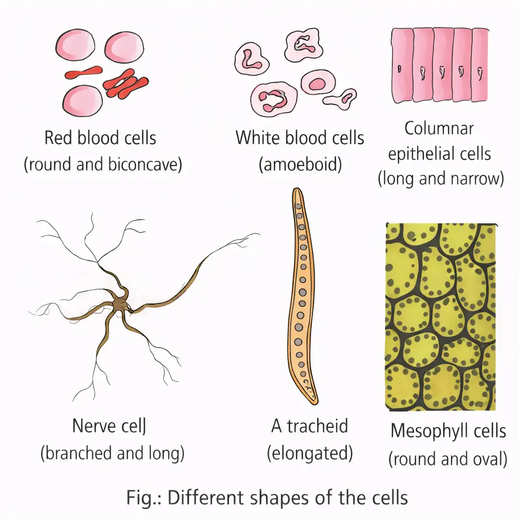
- Cell size varies: Mycoplasmas are smallest (~0.3 µm), bacteria 3-5 µm, human red blood cells ~7 µm, nerve cells are very long.
- Cell shapes vary: disc-like, polygonal, columnar, cuboid, thread-like, or irregular, depending on function.
Memory Aids
- Eukaryotic = "Eu" means true nucleus present.
- Prokaryotic = "Pro" means before nucleus (no membrane-bound nucleus).
- Ribosome = "Organelle within organelle" (found inside mitochondria and chloroplasts).
- Cell shape matches function: e.g., nerve cells are long for signal transmission.
Quick Checkpoints
- What defines a eukaryotic cell?
- Which organelles are membrane-bound?
- Where are ribosomes found inside the cell?
- How does cell shape relate to its function?
- What is the role of centrioles?
Prokaryotic Cells
Characteristics and Types
- Represented by bacteria, blue-green algae, mycoplasma, and PPLO.
- Smaller and multiply faster than eukaryotic cells.
- Shapes: Bacillus (rod), Coccus (spherical), Vibrio (comma), Spirillum (spiral).
- Cell wall present except in mycoplasma.
- Cytoplasm lacks membrane-bound nucleus; DNA is naked and circular.
- Plasmids: small circular DNA outside genomic DNA, confer traits like antibiotic resistance.
- No organelles except 70S ribosomes.
- Mesosome: infoldings of plasma membrane, unique to prokaryotes.
Memory Aids
- Prokaryotes = No nucleus, No organelles except ribosomes.
- Bacterial shapes: B-C-V-S (Bacillus, Coccus, Vibrio, Spirillum).
- Plasmids = Extra DNA for special traits.
Quick Checkpoints
- What shapes do bacteria exhibit?
- Which prokaryotes lack a cell wall?
- What is the role of plasmids?
- What is a mesosome?
Cell Envelope and Modifications
Structure and Types
- Cell envelope: glycocalyx, cell wall, plasma membrane.
- Glycocalyx: slime layer (loose) or capsule (thick).
- Cell wall: maintains shape, prevents bursting.
- Plasma membrane: selectively permeable, similar to eukaryotes.
- Gram staining divides bacteria into Gram-positive (retain stain) and Gram-negative (do not retain).
- Mesosome: plasma membrane infoldings as vesicles, tubules, lamellae.
- Mesosome functions: cell wall formation, DNA replication, distribution, respiration, secretion, surface area increase.
- Cyanobacteria have chromatophores with pigments.
Memory Aids
- Glycocalyx: Slime = loose, Capsule = tough.
- Gram-positive = purple stain, Gram-negative = pink/red.
- Mesosome = membrane infoldings aiding multiple cell functions.
Quick Checkpoints
- What layers form the cell envelope?
- Difference between slime layer and capsule?
- Functions of mesosome?
- How are bacteria classified by Gram stain?
Bacterial Surface Structures
Motility and Attachment
- Flagella: thin filaments for motility; parts: filament (longest), hook, basal body.
- Number and arrangement of flagella vary among bacteria.
- Pili: tubular protein structures, not for motility.
- Fimbriae: small bristle-like fibers, help attachment to surfaces and host tissues.
Memory Aids
- Flagella = movement; Pili and fimbriae = attachment.
- Flagellum parts: F-H-B (Filament, Hook, Basal body).
Quick Checkpoints
- What are the parts of bacterial flagellum?
- Which structures aid bacterial attachment?
- Do pili help in motility?
Ribosomes and Inclusion Bodies
Structure and Function
- Prokaryotic ribosomes are 70S (50S + 30S), size 15-20 nm.
- Site of protein synthesis.
- Polyribosomes (polysomes): multiple ribosomes on one mRNA for simultaneous translation.
- Inclusion bodies: reserve materials like phosphate, cyanophycean, glycogen granules; not membrane-bound.
- Gas vacuoles in photosynthetic bacteria for buoyancy.
Memory Aids
- 70S ribosomes = Prokaryotes.
- Polysomes = multiple ribosomes on mRNA.
- Inclusion bodies = storage granules.
Quick Checkpoints
- What is the size and type of prokaryotic ribosomes?
- What are polysomes?
- What types of inclusion bodies exist?
- Function of gas vacuoles?
Eukaryotic Cells
Structure and Components
- Eukaryotes include protists, plants, animals, and fungi.
- Cells have a well-organized nucleus with nuclear envelope.
- Contain several membrane-bound organelles.
- Genetic material is organized into chromosomes and chromatin.
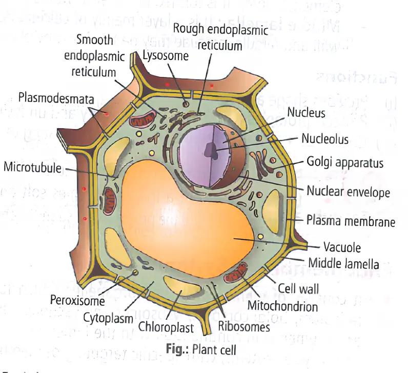
- Cell wall: outer layer providing shape and protection.
- Plasma membrane: controls entry and exit of substances.
- Nucleus: contains genetic material, controls activities.
- Nuclear envelope: protects nucleus.
- Nucleolus: site of ribosome synthesis.
- Cytoplasm: jelly-like substance housing organelles.
- Chloroplasts: site of photosynthesis.
- Mitochondrion: produces cellular energy.
- Vacuole: stores water and nutrients.
- Golgi apparatus: packages and transports proteins.
- Ribosomes: protein synthesis.
- Endoplasmic reticulum (rough and smooth): protein and lipid synthesis and transport.
- Lysosomes: waste breakdown.
- Peroxisomes: detoxify harmful substances.
- Microtubules: maintain cell shape.
- Plasmodesmata: channels for cell communication.
- Middle lamella: glues plant cells together.
Memory Aids
- “Nucleus controls cell activities.”
- “Chloroplasts make food via photosynthesis.”
- “Mitochondria are the cell’s powerhouses.”
- “Golgi apparatus packages proteins.”
Quick Checkpoints
- What organelle controls cell activities?
- Where does photosynthesis occur?
- Which organelle produces energy?
- What is the function of Golgi apparatus?
Cell Membrane
Structure and Composition
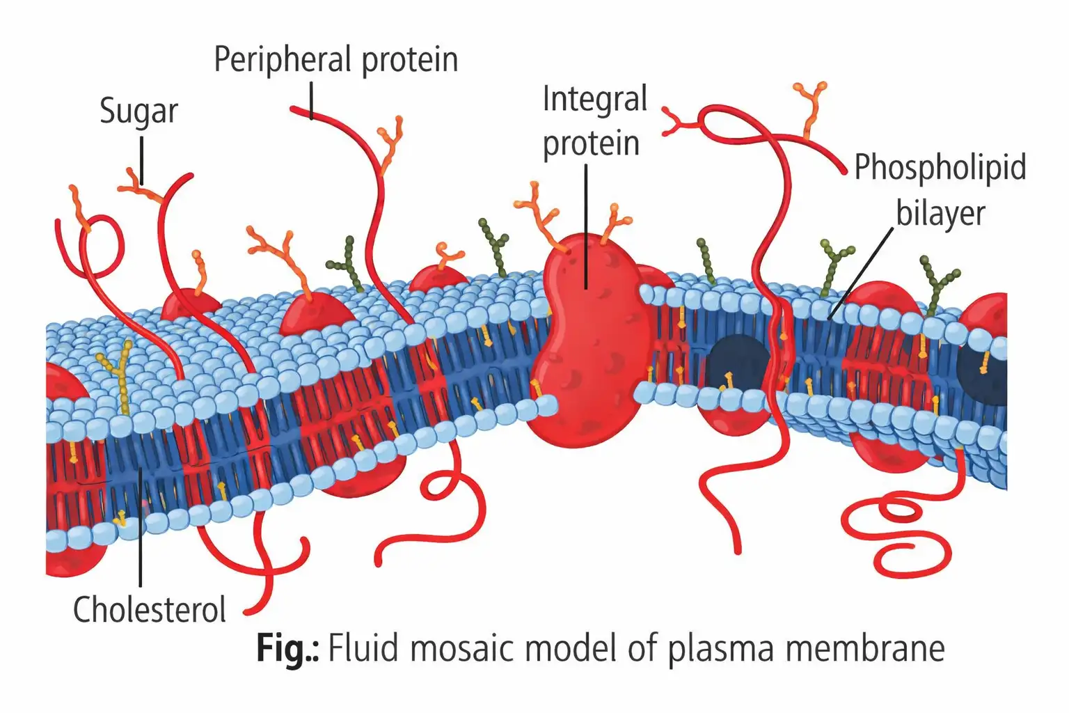
- Composed mainly of phospholipids and proteins; also contains cholesterol.
- Lipids arranged with polar heads outward and hydrophobic tails inward.
- Contains lipids (20-79%), proteins (20-70%), carbohydrates (1-5%), and water (20%).
- Proteins classified as integral (embedded) or peripheral (surface).
- Fluid mosaic model: proteins float in a lipid bilayer, allowing lateral movement.
Functions
- Selective permeability controls molecule transport.
- Passive transport: diffusion and osmosis without energy.
- Active transport: energy-dependent movement against concentration gradient (e.g., Na+/K+ pump).
Memory Aids
- “Fluid mosaic: proteins float in lipid sea.”
- “Integral proteins embedded; peripheral on surface.”
- “Passive transport needs no energy; active transport does.”
Quick Checkpoints
- What are the main components of the cell membrane?
- Define fluid mosaic model.
- Difference between passive and active transport?
- Role of cholesterol in membrane?
Cell Wall
Structure and Composition
- Non-living, rigid, protective outer covering in plants, fungi, some protists.
- Algal walls: cellulose, galactans, mannans, calcium carbonate.
- Plant walls: cellulose, hemicellulose, pectins, proteins.
- Three parts: primary wall (young cells), middle lamella (calcium pectate, glues cells), secondary wall (mature cells).
- Plasmodesmata connect cytoplasm of adjacent cells.
Functions
- Provides shape and rigidity.
- Protects protoplasm from injury and macromolecules.
- Prevents bursting by counteracting osmotic pressure.
Memory Aids
- “Microfibrils are structural elements.”
- “Middle lamella dissolves during fruit ripening.”
- “Plasmodesmata discovered by Strasburger.”
Quick Checkpoints
- What are the three parts of the cell wall?
- Function of middle lamella?
- Role of plasmodesmata?
Endomembrane System
Components and Functions
- Includes endoplasmic reticulum, Golgi complex, lysosomes, vacuoles.
- Nuclear envelope lumen continuous with ER lumen.
- Functions in processing, sorting, and recycling proteins.
Memory Aids
- “Endomembrane system processes and sorts proteins.”
Quick Checkpoints
- Which organelles form the endomembrane system?
- How is nuclear envelope related to ER?
Endoplasmic Reticulum
Structure and Types
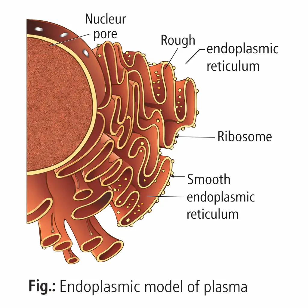
- Network of tubular structures dividing cytoplasm into luminal (inside ER) and extra luminal (cytoplasm) compartments.
- Composed of cisternae (protein synthesis), tubules (lipid synthesis), and vesicles.
- Two types: Rough ER (RER) with ribosomes, Smooth ER (SER) without ribosomes.
Functions
- RER: protein synthesis and secretion.
- RER: glycosylation and lysosome enzyme precursor formation.
- SER: lipid and steroid hormone synthesis.
- SER: detoxification of drugs and xenobiotics.
Memory Aids
- “RER has ribosomes; SER does not.”
- “SER detoxifies and synthesizes lipids.”
Quick Checkpoints
- Difference between RER and SER?
- Role of ER in protein synthesis?
- Function of SER in animal cells?
Golgi Apparatus
Structure and Occurrence
- Discovered by Camillo Golgi in 1898.
- Present in all eukaryotic cells except male gametes of bryophytes, mammalian RBCs, sieve tubes, fungi cells.
- Animal cells: single or connected complex; plant cells: unconnected dictyosomes.
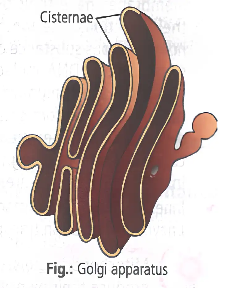
- Composed of cisternae, vesicles, tubules, and vacuoles.
- Cisternae are flat sacs stacked parallel with distinct cis (forming) and trans (maturing) faces.
Functions
- Packages and stores materials from ER.
- Modifies proteins before release.
- Forms acrosomes during spermatogenesis.
- Glycosylates proteins and lipids for cell recognition and adhesion.
Memory Aids
- “Golgi acts like a post office of the cell.”
- “Cis face receives; trans face ships.”
Quick Checkpoints
- What are the parts of Golgi apparatus?
- Function of cis and trans faces?
- Role in protein modification?
Lysosomes
Structure and Composition
- Discovered by Christian de Duve in 1955.
- Single membrane-bound vesicles containing hydrolytic enzymes (lipases, proteases, carbohydrases).
- Formed by Golgi apparatus packaging.
- Known as suicidal bags due to digestive enzymes.
- Present in all animal cells except RBCs; vacuoles perform similar function in plants and fungi.
Types
- Primary lysosomes, secondary lysosomes, residual bodies, autophagic vacuoles.
Memory Aids
- “Lysosomes digest waste and old organelles.”
- “Called suicidal bags due to enzymes.”
Quick Checkpoints
- What enzymes do lysosomes contain?
- Why are lysosomes called suicidal bags?
- Types of lysosomes?
Vacuoles
Structure and Types
- Membrane-bound spaces in cytoplasm containing water, sap, excretory products.
- Bounded by tonoplast membrane.
- Plant vacuoles can occupy up to 90% of cell volume.
- Types: sap vacuole, food vacuole, contractile vacuole, air vacuole.
- Contractile vacuole in Amoeba regulates osmoregulation and excretion.
- Food vacuoles formed by engulfing food in protists.
- Tonoplast controls ion passage, maintains osmotic pressure and turgidity.
Memory Aids
- “Tonoplast surrounds vacuole.”
- “Contractile vacuole controls water balance in protists.”
Quick Checkpoints
- What is tonoplast?
- Functions of vacuoles?
- Types of vacuoles?
Mitochondria
Discovery and Occurrence
- Observed by Kolliker (1857); named by Benda (1898).
- Present in aerobic eukaryotes; absent in prokaryotes and mature RBCs.
- Known as powerhouses of the cell for energy production.
Structure
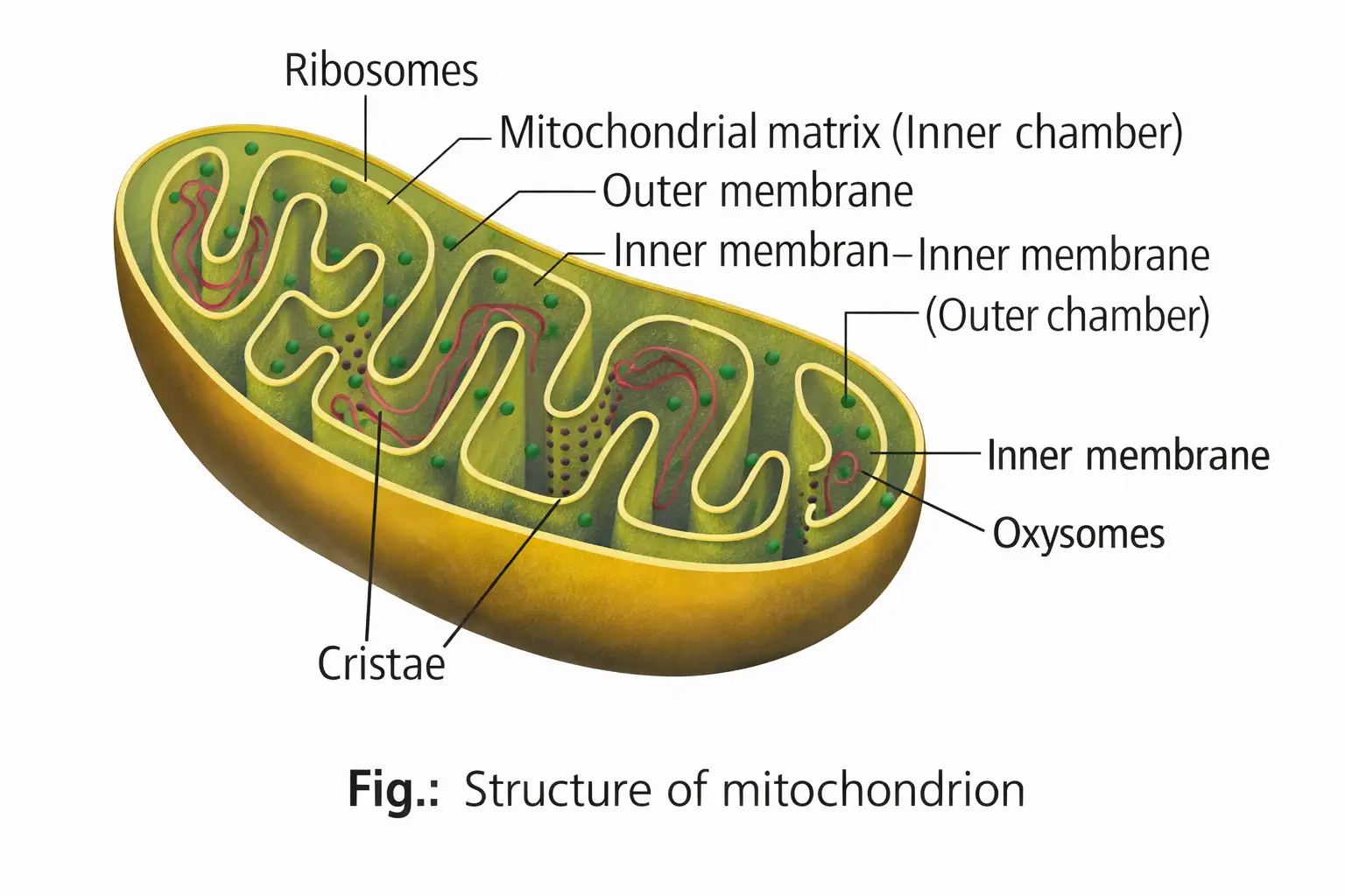
- Cylindrical, double-membraned organelles (1-4.1 μm length, 0.2-1 μm diameter).
- Outer membrane: smooth, continuous boundary.
- Inner membrane: folded into cristae to increase surface area.
- Matrix: dense fluid inside inner membrane containing circular DNA, RNA, 70S ribosomes, enzymes.
- Inner membrane and cristae have elementary particles (F0-F1 particles) with electron transport enzymes.
- Divide by fission.
Memory Aids
- “Cristae increase surface area for respiration.”
- “Mitochondria have their own DNA and ribosomes.”
- “Divide by fission like bacteria.”
Quick Checkpoints
- Describe mitochondrial structure.
- Function of cristae?
- Why are mitochondria semi-autonomous?
Plastids
Types and Functions
- Found in plant cells and euglenoids; contain pigments.
- Three types based on pigments: leucoplasts (colorless, store food), chloroplasts (green, photosynthesis), chromoplasts (yellow/red, pigment storage).
- Leucoplasts: amyloplasts (starch), elaioplasts (fat/oil), aleuroplasts (protein).
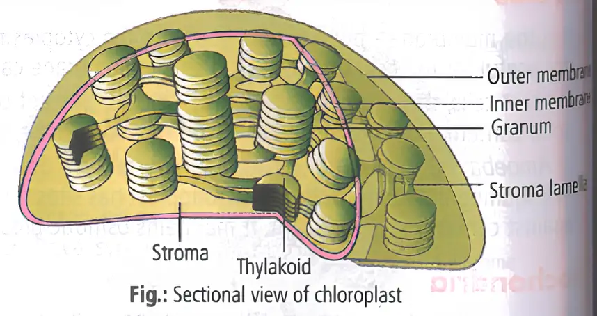
- Chloroplasts: lens-shaped, double membrane, contain stroma and thylakoids arranged in grana.
- Chromoplasts: carotenoid pigments, color flowers/fruits, aid pollination and seed dispersal.
- Chloroplasts have DNA, 70S ribosomes, and enzymes; semi-autonomous.
Memory Aids
- “Chloroplasts for photosynthesis.”
- “Leucoplasts store food.”
- “Chromoplasts give color to plants.”
Quick Checkpoints
- Types of plastids and their functions?
- Structure of chloroplast?
- Why are chloroplasts semi-autonomous?
Ribosomes
Structure and Function
- Discovered by George Palade (1953).
- Composed of RNA and proteins; no membrane.
- Eukaryotic ribosomes are 80S (60S + 40S subunits); prokaryotic are 70S (50S + 30S).
- Sites of protein synthesis; act as protein factories.
Memory Aids
- “80S in eukaryotes, 70S in prokaryotes.”
- “Two subunits form functional ribosome.”
Quick Checkpoints
- What are ribosomes made of?
- Difference between eukaryotic and prokaryotic ribosomes?
- Function of ribosomes?
Cytoskeleton
Structure and Types
- Network of protein filaments in cytoplasm.
- Maintains cell shape, organelle distribution, intracellular transport, and movement.
- Types: microfilaments, microtubules, intermediate filaments.
Memory Aids
- “Cytoskeleton supports and moves the cell.”
- “Three filament types for structure and transport.”
Quick Checkpoints
- What are the three types of cytoskeletal elements?
- Functions of cytoskeleton?
Cilia and Flagella
Structure and Function
- Hair-like outgrowths of plasma membrane.
- Cilia: short, oar-like movement of cell or fluid.
- Flagella: longer, responsible for cell movement.
- Prokaryotic flagella structurally different from eukaryotic.
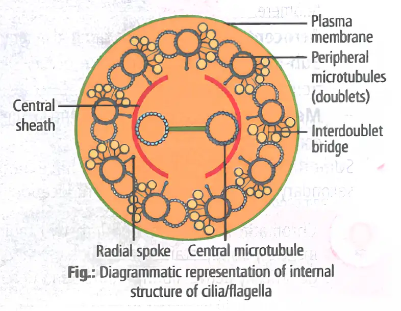
- Core (axoneme) has 9+2 microtubule arrangement: nine pairs around two central microtubules.
- Connected by radial spokes and interdoublet linkers; arise from basal bodies.
Memory Aids
- “9+2 arrangement in axoneme.”
- “Cilia short, flagella long.”
- “Basal bodies anchor cilia and flagella.”
Quick Checkpoints
- Describe axoneme structure.
- Difference between cilia and flagella?
- Origin of basal bodies?
Centrosome and Centrioles
Structure and Occurrence
- Centrosome contains two centrioles arranged perpendicularly.
- Centrioles present in animal cells and motile plant cells (e.g., zoospores, sperm cells).
- Centrioles have nine triplet microtubules arranged in a cartwheel pattern.
- Central hub connected to triplets by radial spokes.
- Centrioles form basal bodies of cilia/flagella and spindle fibers during cell division.
Memory Aids
- “Centrioles have 9 triplet microtubules.”
- “Form basal bodies and spindle fibers.”
Quick Checkpoints
- Structure of centrioles?
- Function of centrosome?
- Where are centrioles found?
Nucleus
Structure and Components
- Discovered by Robert Brown (1831).
- Largest organelle with double membrane.
- Contains chromatin, nucleoplasm, nucleoli.
- Nuclear envelope has pores for RNA and protein transport.
- Outer membrane continuous with ER; bears ribosomes.
- Usually one nucleus per cell; some binucleate or multinucleate cells exist.
- Chromatin: DNA + histones + non-histone proteins + RNA.
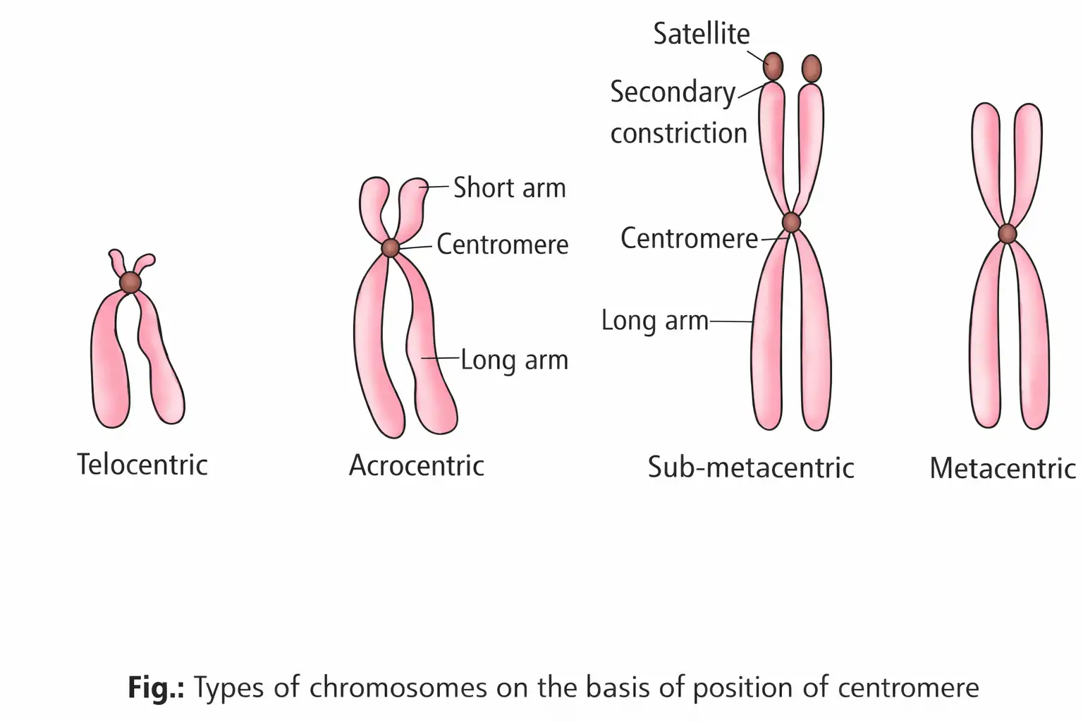
- Chromosomes have centromere and kinetochores.
Chromosome Types
- Telocentric: centromere at terminal end.
- Acrocentric: centromere near end.
- Sub-metacentric: centromere slightly off center.
- Metacentric: centromere in middle.
- Some chromosomes have satellites (secondary constrictions).
Memory Aids
- “Chromatin: euchromatin (active), heterochromatin (inactive).”
- “Nucleolus synthesizes rRNA.”
- “Nuclear pores allow molecule exchange.”
Quick Checkpoints
- What is chromatin made of?
- Types of chromosomes based on centromere?
- Function of nucleolus?
Microbodies
Structure and Function
- Membrane-bound vesicles containing enzymes.
- Present in both plant and animal cells.
Memory Aids
- “Microbodies contain various enzymes.”
Quick Checkpoints
- What are microbodies?
- Where are microbodies found?
Plant Cell v/s Animal Cell
Differences
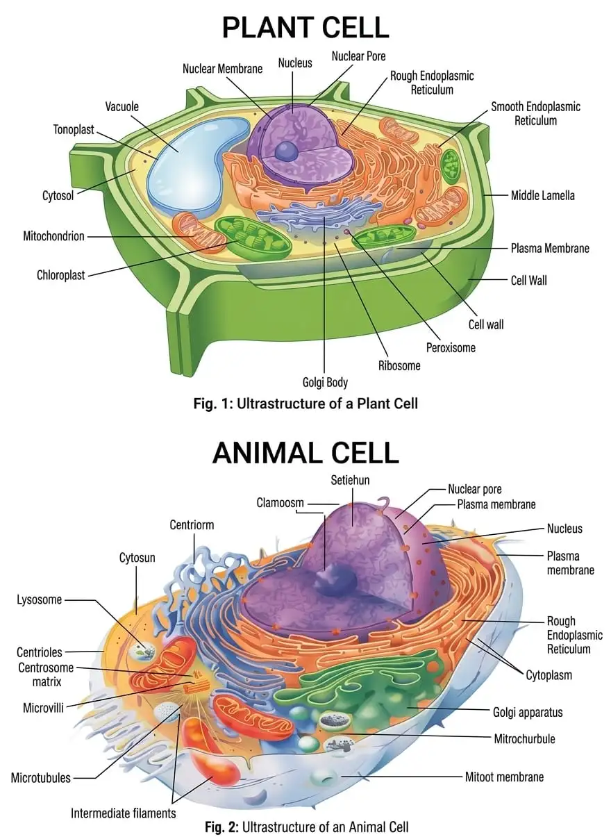
- Plant cells have cell wall, plastids, large central vacuole; absent in animal cells.
- Animal cells have centrioles; absent in most plant cells.
Memory Aids
- “Plant cells: wall, plastids, big vacuole.”
- “Animal cells: centrioles present.”
Quick Checkpoints
- Key structural differences between plant and animal cells?
- Functions of plastids and vacuoles in plants?
- Role of centrioles in animal cells?
NEET Botany — ALL CHAPTERS
1
The Living World
2
Plant Kingdom
3
Morphology of Flowering Plants
4
Anatomy of Flowering Plants
5
The Unit of Life
6
Photosynthesis in Higher Plants
7
Respiration in Plants
8
Plant Growth and Development
9
Sexual Reproduction in Flowering Plants
10
Principles of Inheritance and Variation
11
Molecular Basis of Inheritance
12
Biotechnology :Principles and Processes
13
Biotechnology and its Applications