NEET Ug . NEET Botany . Botany . Molecular Basis of Inheritance
Chapter 11: Molecular Basis of Inheritance
Ch 11
NEET Botany
NEET Ug
Introduction
Overview
- At Mendel's time, inheritance factors were unknown.
- DNA and RNA are nucleic acids made of nucleotides.
- DNA is genetic material in most organisms.
- RNA acts as genetic material in some viruses and functions mainly as a messenger, adapter, structural, or catalytic molecule.
Memory Aids
- DNA = Genetic blueprint in most life forms.
- RNA = Messenger and catalyst in cells.
Quick Checkpoints
- What are the two main nucleic acids?
- Which nucleic acid acts as genetic material in most organisms?
The DNA
Structure and Composition
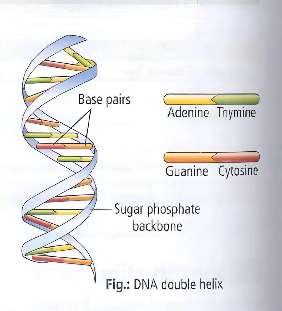
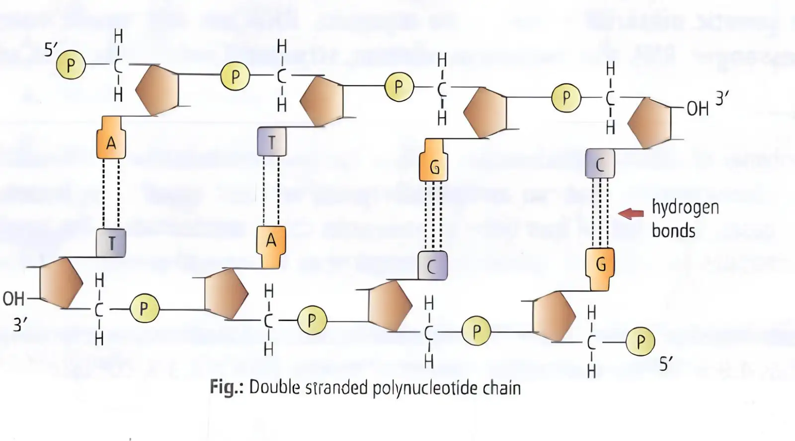
- DNA is a long polymer of deoxyribonucleotides forming two complementary strands running antiparallel.
- Strands held by hydrogen bonds between opposite nitrogen bases.
- Four types of nucleotides form DNA.
- DNA length is measured by number of nucleotides or base pairs (bp).
- Examples: Φ x174 (5386 nucleotides), lambda phage (48502 bp), Escherichia coli (4.6 million bp), human haploid DNA (3.3 billion bp).
Memory Aids
- DNA strands run antiparallel and complement each other.
- Base pairs define DNA length and organism specificity.
Quick Checkpoints
- What is the orientation of DNA strands?
- How is DNA length defined?
Structure of Polynucleotide Chain
Components and Linkages
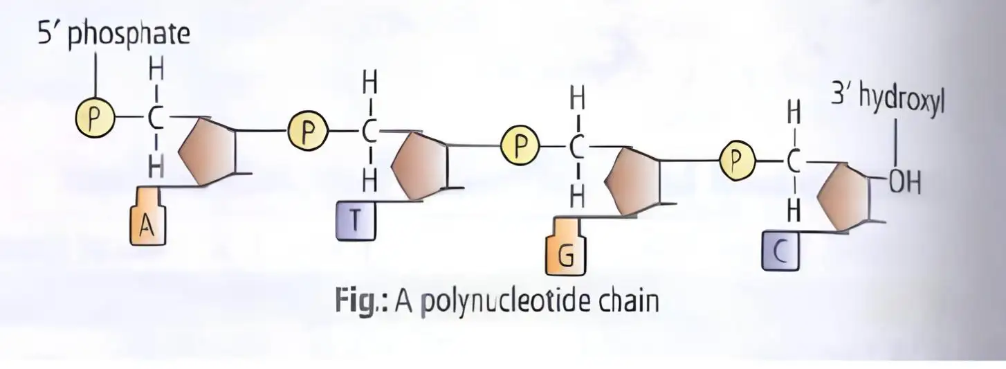
- Nucleotide = nitrogenous base + pentose sugar + phosphate group.
- Purines: adenine, guanine; Pyrimidines: cytosine, uracil (RNA), thymine (DNA).
- Cytosine common to DNA and RNA; uracil replaces thymine in RNA.
- Base linked to 1′C sugar via N-glycosidic linkage forming nucleoside.
- Phosphate linked to 5′-OH of nucleoside via phosphodiester bond forms nucleotide.
- Nucleotides linked 3′–5′ phosphodiester form dinucleotides and polynucleotide chains.
- RNA nucleotides have extra –OH at 2′ position; DNA identified by Friedrich Miescher in 1869 as 'Nuclein'.
Memory Aids
- Nucleotide = Base + Sugar + Phosphate.
- RNA has uracil; DNA has thymine.
- Phosphodiester bonds link nucleotides 3′ to 5′.
Quick Checkpoints
- What are the three parts of a nucleotide?
- Which base replaces thymine in RNA?
- What type of bond links nucleotides?
Structure of DNA
Double Helix Model
- Proposed by James Watson and Francis Crick in 1953, based on X-ray data by Wilkins and Franklin.
- Chargaff's rules: purines = pyrimidines (A + G = T + C), A = T, G = C.
- Base ratio A + T / G + C varies by species but constant within species.
- Each DNA strand acts as template for identical daughter DNA.
- Features:
- Two polynucleotide chains with sugar-phosphate backbone; bases inside.
- Chains are antiparallel (5′→3′ and 3′→5′).
- Bases paired by hydrogen bonds: A-T (2 bonds), G-C (3 bonds); purine pairs with pyrimidine.
- Right-handed double helix with 3.4 nm pitch and ~10 bp per turn.
- Base pairs stack for stability.
Memory Aids
- DNA strands are antiparallel and complementary.
- A pairs with T; G pairs with C.
- Double helix is right-handed with 10 bp per turn.
Quick Checkpoints
- Who proposed the DNA double helix model?
- What are Chargaff's base pairing rules?
- Describe the polarity of DNA strands.
Central Dogma
Genetic Information Flow
- Proposed by Francis Crick.
- Genetic information flows: DNA → RNA → Protein.
- Steps: DNA replication, transcription (DNA to mRNA), translation (mRNA to protein).
- In some viruses, reverse flow occurs: RNA → DNA (reverse transcription).


Memory Aids
- DNA → RNA → Protein is the central dogma.
- Reverse transcription occurs in some viruses.
Quick Checkpoints
- What is the direction of genetic information flow?
- What is reverse transcription?
Packaging of DNA Helix
DNA Length and Compaction
- DNA length = number of base pairs × distance between base pairs.
- Typical mammalian cell DNA length ≈ 2.2 meters.
- DNA is compacted to fit in small nucleus.
Memory Aids
- DNA is very long but tightly packed.
Quick Checkpoints
- How is DNA length calculated?
- Why is DNA packaging necessary?
DNA Packaging in Prokaryotes
Supercoiling and Nucleoid
- DNA is supercoiled with help of RNAs and non-histone proteins like polyamines.
- Compacted DNA mass is called nucleoid.
Memory Aids
- Prokaryotic DNA is supercoiled and forms nucleoid.
Quick Checkpoints
- What is nucleoid?
- How is prokaryotic DNA compacted?
DNA Packaging in Eukaryotes
Histones and Nucleosomes
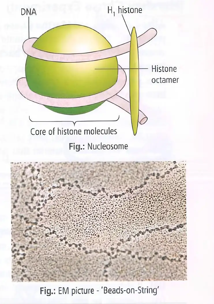
- DNA wraps around histones (lysine and arginine rich proteins).
- Histones form octamer (eight molecules) called histone octamer.
- DNA + histone octamer = nucleosome.
- Five histone types: H1, H2A, H2B, H3, H4; H2A, H2B, H3, H4 form octamer.
- Typical nucleosome contains ~200 bp DNA.
- Nucleosomes form chromatin, visible as thread-like bodies in nucleus.
- Chromatin appears as 'beads-on-string' under electron microscope.
- Chromatin fibers coil and condense into chromosomes during metaphase.
- Non-histone chromosomal proteins (NHC) assist higher-level packaging.
- Chromatin types: euchromatin (loosely packed, active), heterochromatin (densely packed, inactive).
- DNA between nucleosomes is linker DNA or inerbead DNA, bearing H1 histone (plugging and marker protein).
- Nucleosome + linker DNA = chromatosome.
Memory Aids
- Histones package DNA into nucleosomes.
- Chromatin = nucleosomes + linker DNA.
- Euchromatin = active; heterochromatin = inactive.
Quick Checkpoints
- What proteins help package eukaryotic DNA?
- Define nucleosome and chromatosome.
- Difference between euchromatin and heterochromatin?
Transforming Principle
Key Findings
- 1928: Frederick Griffith studied Streptococcus pneumoniae with two strains: S-type (virulent, smooth colonies) and R-type (non-virulent, rough colonies).
- Heat-killed S strain alone did not kill mice; live R strain alone did not kill mice.
- Mix of heat-killed S strain and live R strain killed mice, showing transformation of R to virulent S type.
- Conclusion: A 'transforming principle' from dead S strain transferred genetic material to R strain, enabling virulence.
Memory Aids
- Griffith’s experiment: Dead S + Live R = Dead mice (transformation).
- Transforming principle = genetic material transfer.
Quick Checkpoints
- What are the two strains of Streptococcus pneumoniae?
- What did the heat-killed S strain demonstrate?
- What is the significance of the transforming principle?
Biochemical Characterisation of Transforming Principle (Avery Experiment)
Key Findings
- Oswald Avery, Colin MacLeod, and Maclyn McCarty (1933-44) purified proteins, DNA, and RNA from heat-killed S cells.
- Only DNA caused transformation of live R cells into S cells.
- Protease and RNase treatments did not stop transformation; DNase treatment did.
- Conclusion: DNA is the hereditary material, not protein or RNA.
Memory Aids
- DNase stops transformation → DNA is genetic material.
- Protease and RNase do not affect transformation.
Quick Checkpoints
- Which enzyme inhibited transformation?
- What was the role of protease and RNase?
- What did Avery’s experiment prove about genetic material?
DNA is the Genetic Material (Hershey Chase Experiment)
Key Findings
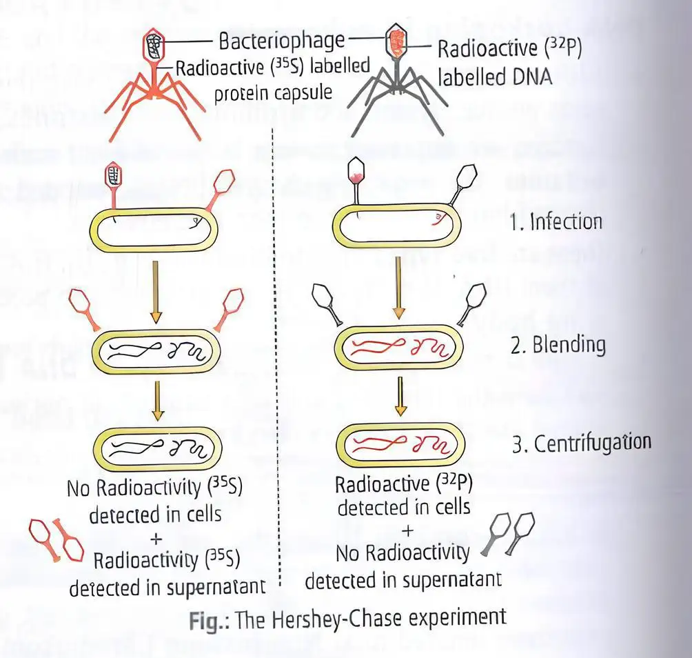
- Alfred Hershey and Martha Chase (1952) used bacteriophages to prove DNA is genetic material.
- Phages labeled with radioactive phosphorus (32P) in DNA and radioactive sulfur (35S) in protein.
- Only radioactive DNA entered Escherichia coli cells, not protein.
- Conclusion: DNA carries genetic information, not protein.
Memory Aids
- 32P labels DNA; 35S labels protein.
- DNA enters bacteria; protein does not.
- DNA = genetic material.
Quick Checkpoints
- What isotopes were used to label DNA and protein?
- Which component entered bacterial cells?
- What was the conclusion of the Hershey-Chase experiment?
Properties of Genetic Material (DNA versus RNA)
Key Properties
- Some viruses use RNA as genetic material (e.g., Tobacco Mosaic virus, QB bacteriophage).
- Genetic material must: replicate, be chemically and structurally stable, allow mutation, and express Mendelian characters.
- DNA and RNA can replicate due to base pairing; proteins cannot.
- DNA is chemically more stable than RNA due to absence of reactive 2'-OH group and presence of thymine instead of uracil.
- RNA is reactive, catalytic, and degrades easily.
- RNA mutates faster; RNA viruses evolve faster.
- RNA directly codes proteins; DNA depends on RNA for protein synthesis.
- DNA is preferred for genetic information storage; RNA for transmission.
Memory Aids
- DNA = stable, stores info; RNA = reactive, transmits info.
- Thymine in DNA adds stability; uracil in RNA less stable.
- RNA mutates faster → faster evolution.
Quick Checkpoints
- What are the criteria for genetic material?
- Why is DNA more stable than RNA?
- How do RNA and DNA differ in mutation rates and roles?
RNA World
RNA as First Genetic Material
- RNA was the first genetic material supporting essential life processes like metabolism, translation, and splicing.
- RNA acted both as genetic material and catalyst but was chemically reactive and unstable.
- DNA evolved from RNA with chemical changes making it more stable.
- DNA is double-stranded with complementary strands and repair mechanisms to resist changes.
Types of Non-Genetic RNA
- Based on size and function, three main non-genetic RNAs: mRNA (~3.5%), tRNA (~15%), rRNA (~80%).
- mRNA: Transcribed from genes, serves as template for protein synthesis by ribosomes.
- tRNA: Also called soluble, acceptor, or adapter RNA; has amino acid binding site opposite anticodon.
- rRNA: Structural component of ribosomes, synthesized in nucleolus.
Memory Aids
- RNA = First genetic material and catalyst.
- DNA = Stable, double-stranded, repair-capable.
- mRNA = Messenger, carries code.
- tRNA = Transfer, carries amino acids.
- rRNA = Ribosome, protein factory.
Quick Checkpoints
- What roles did RNA play in early life?
- How is DNA chemically more stable than RNA?
- What are the main types of non-genetic RNA and their functions?
- Where is rRNA synthesized?
Replication
Process and Mechanism
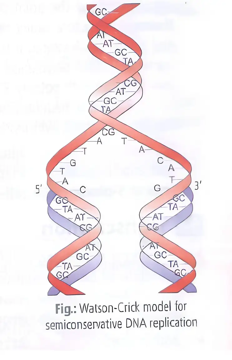
- DNA replication is the process of making an identical copy of double-stranded DNA using the original DNA as a template.
- Watson and Crick proposed semiconservative replication: each new DNA molecule has one parental and one newly synthesized strand.
- Replication involves unwinding the double helix and synthesizing complementary strands.
Memory Aids
- "Semi" means half old, half new strands in each DNA molecule.
- Complementary base pairing: A pairs with T, G pairs with C.
Quick Checkpoints
- What is semiconservative replication?
- Which strands serve as templates during replication?
- How does base pairing ensure accurate copying?
The Experimental Proof (Meselson and Stahl Experiment)
Experiment and Findings
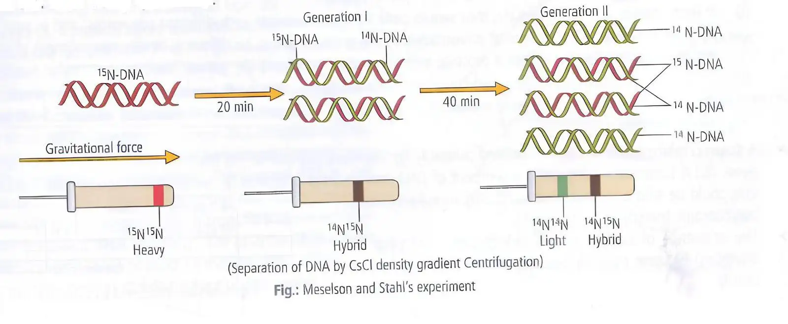
- Meselson and Stahl (1958) proved DNA replicates semi-conservatively using Escherichia coli and nitrogen isotopes.
- DNA labeled with heavy nitrogen (15N) was grown in light nitrogen (14N) medium.
- After one generation, DNA density was intermediate, showing hybrid strands with one old and one new strand.
- Subsequent generations showed a mix of hybrid and light DNA, confirming semiconservative replication.
- Taylor et al. confirmed semi-conservative replication in chromosomes of Vicia faba using radioactive thymidine.
Memory Aids
- Heavy nitrogen (15N) labels old DNA strands.
- Hybrid DNA has one heavy and one light strand.
- Density gradient centrifugation separates DNA by weight.
Quick Checkpoints
- What isotope was used to label DNA in the experiment?
- How does DNA density change after replication in light nitrogen?
- What does the presence of hybrid DNA indicate?
The Machinery and the Enzymes
Enzymes and Process
- DNA replication is energy-intensive and requires enzymes called catalysts.
- Main enzyme: DNA-dependent DNA polymerase synthesizes new DNA strands using a DNA template.
- Replication speed in E. coli is about 2000 base pairs per second.
- Energy comes from breakdown of deoxyribonucleotide triphosphates.
- Helicase unwinds DNA at origin of replication (Ori site).
- Single-stranded binding proteins stabilize separated strands.
- Topoisomerases relieve supercoiling by nicking and resealing DNA strands.
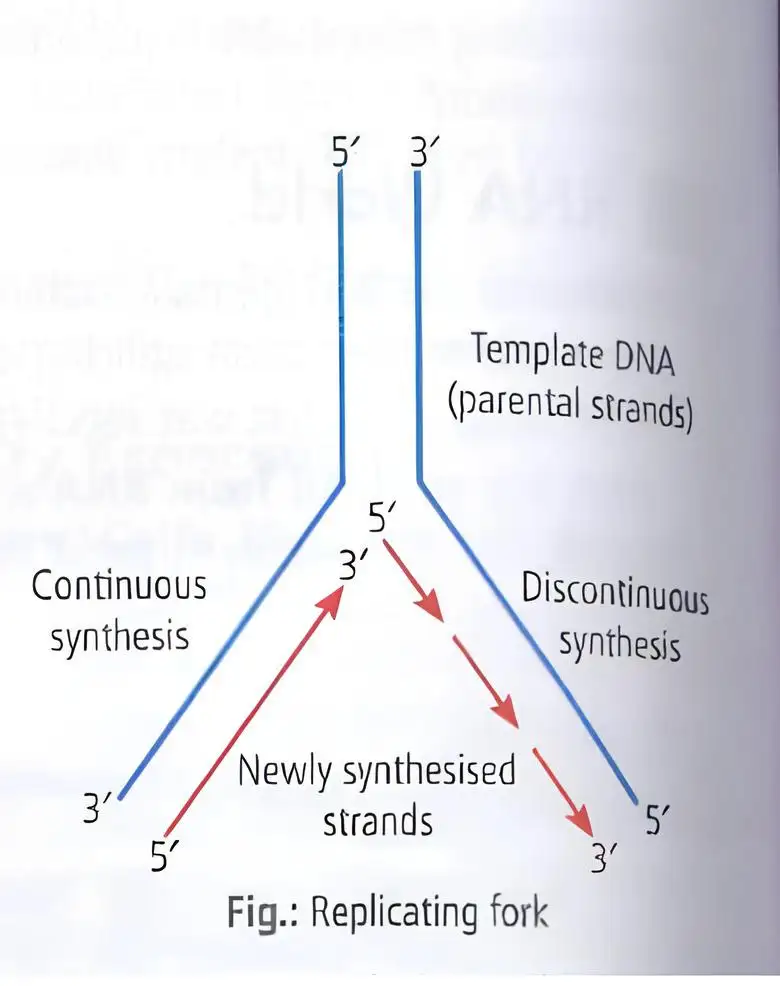
- Replication fork is Y-shaped where DNA strands separate and replication occurs.
- RNA primer initiates new DNA strand synthesis; DNA polymerase adds nucleotides 5' to 3'.
- Leading strand replicates continuously (template 3' to 5').
- Lagging strand replicates discontinuously in Okazaki fragments (template 5' to 3').
- DNA ligase joins Okazaki fragments.
- Replication starts at specific origin sites, not randomly.
- In eukaryotes, replication occurs during S-phase of the cell cycle.
Memory Aids
- Helicase = Unwinds DNA.
- SSB proteins = Stabilize single strands.
- Topoisomerase = Relieves supercoiling.
- DNA polymerase = Synthesizes DNA 5' to 3'.
- RNA primer = Starts DNA synthesis.
- Ligase = Joins DNA fragments.
- Leading strand = Continuous synthesis.
- Lagging strand = Discontinuous synthesis.
Quick Checkpoints
- Which enzyme unwinds the DNA helix?
- What stabilizes single DNA strands during replication?
- Why is RNA primer needed?
- How does replication differ on leading and lagging strands?
- When does DNA replication occur in eukaryotic cells?
Transcription
Process and Mechanism
- Transcription is copying genetic information from one DNA strand into RNA.
- Complementarity governs transcription; adenine pairs with uracil (not thymine).
- Only one DNA strand and a segment are transcribed, unlike replication which duplicates entire DNA.
- Both DNA strands are not copied because:
- If both strands were templates, they would produce different RNA sequences, complicating protein synthesis.
- Simultaneous RNA molecules would be complementary, forming double-stranded RNA, blocking translation.
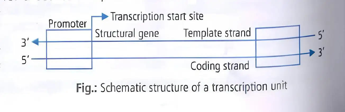
- A transcription unit has three DNA regions: promoter, structural gene, terminator.
- Structural gene can be monocistronic (eukaryotes) or polycistronic (prokaryotes).
- Coding strand sequence matches RNA except thymine is replaced by uracil.
Memory Aids
- Transcription = DNA to RNA copying.
- Only one strand used to avoid conflicting proteins.
- Promoter starts, terminator ends transcription.
- RNA matches coding strand except T → U.
Quick Checkpoints
- Why is only one DNA strand transcribed?
- What are the three regions of a transcription unit?
- How does RNA sequence relate to the coding strand?
Types of RNA and Process of Transcription
RNA Types and Transcription Steps
- Bacteria have three RNA types: mRNA (template), tRNA (amino acid carrier), rRNA (structural/catalytic).
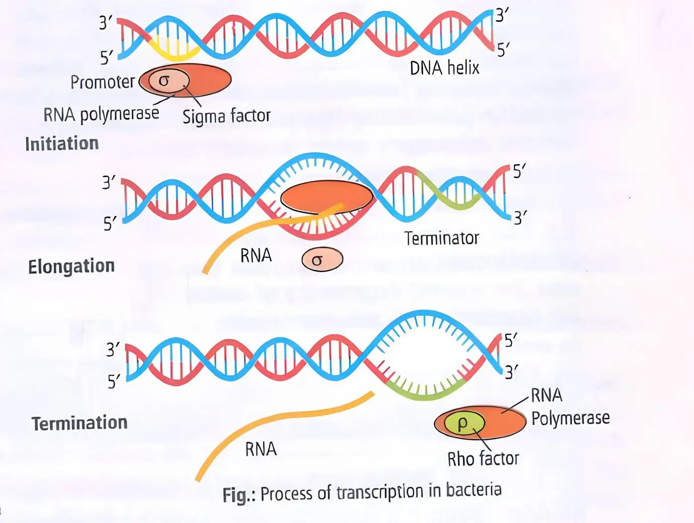
- Transcription enzyme: DNA-dependent RNA polymerase holoenzyme = core enzyme + sigma factor.
- Sigma factor directs RNA polymerase to promoter, enhancing specificity.
- Transcription stages:
- Initiation: RNA polymerase binds promoter (-35 and -10 regions), DNA locally untwists.
- Elongation: Sigma factor leaves after 8-9 nucleotides; RNA polymerase synthesizes RNA 5'→3', unwinding DNA ahead.
- Termination: Rho factor binds RNA, catches polymerase at pause site, unwinds DNA-RNA hybrid, releasing polymerase.
- In bacteria, transcription and translation occur in cytoplasm; mRNA requires no processing.
Memory Aids
- 3 RNA types: mRNA (message), tRNA (transfer), rRNA (ribosome).
- Holoenzyme = core + sigma factor.
- Stages: Initiation (-35/-10), Elongation (5'→3'), Termination (rho factor).
- Bacterial mRNA is ready for translation immediately.
Quick Checkpoints
- What roles do the three RNA types play?
- What is the function of sigma factor?
- Describe the three transcription stages.
- Where does bacterial transcription occur?
Transcription in Eukaryotes
RNA Polymerases and mRNA Processing
- Three RNA polymerases in nucleus:
- RNA polymerase I: transcribes rRNAs (28S, 18S, 5.8S).
- RNA polymerase II: transcribes precursor mRNA (hnRNA).
- RNA polymerase III: transcribes tRNA, 5S rRNA, snRNA.
- Transcription occurs in nucleus; mRNA moves to cytoplasm for translation.
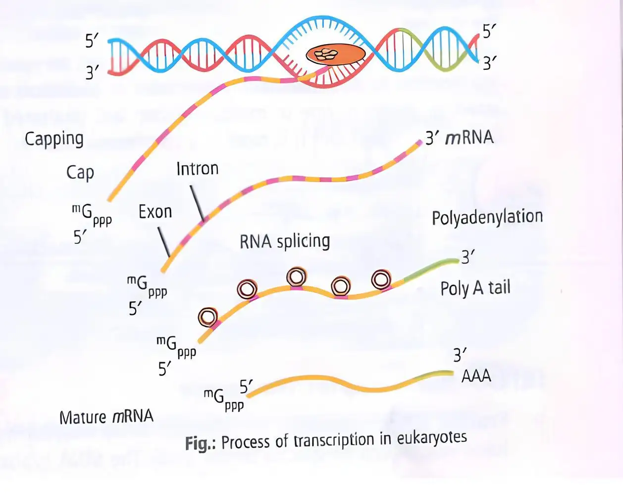
- mRNA maturation includes:
- 5' cap addition (7-methyl guanosine).
- 3' poly-A tail addition (200-300 adenines).
- Removal of introns by RNA splicing.
- Joining of exons by ligase to form mature mRNA.
- Primary transcript called heterogeneous nuclear RNA (hnRNA) or pre-mRNA.
- Splicing shows RNA world dominance.
Memory Aids
- RNA polymerase I = rRNA, II = pre-mRNA, III = tRNA + 5S rRNA + snRNA.
- mRNA processing: cap, tail, splicing.
- hnRNA = pre-mRNA with introns and exons.
- Splicing removes introns, joins exons.
Quick Checkpoints
- Which RNA polymerase transcribes pre-mRNA?
- What modifications occur during mRNA maturation?
- What is hnRNA?
- Why is RNA splicing important?
Genetic Code
Characteristics
- Genetic code is the codon representation decoding RNA into polypeptides.
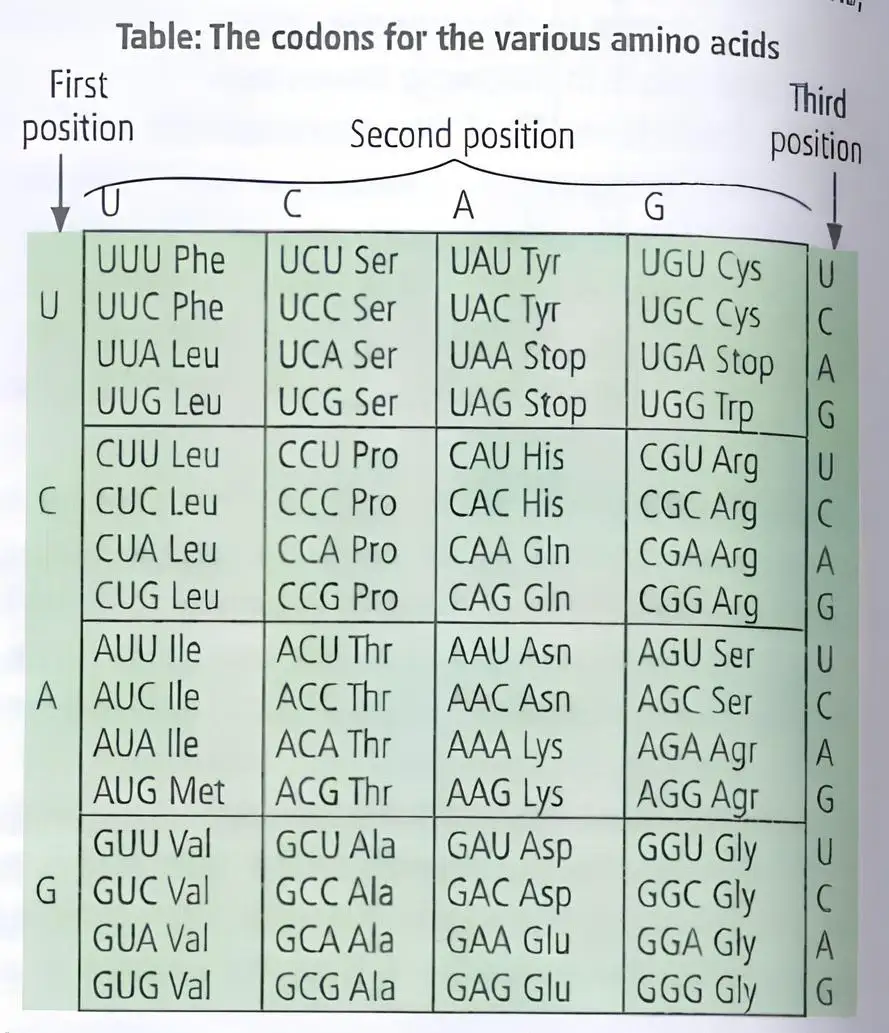
- Code consists of three nucleotides (triplet code) producing 64 codons for 20 amino acids.
- Universal: Same codon codes same amino acid in all organisms (e.g., UUU codes phenylalanine).
- Degeneracy: Multiple codons can code for one amino acid.
- Non-overlapping: Each base is part of only one codon.
- Non-ambiguous: One codon codes for one amino acid, except AUG which can code methionine (start) or valine (internal).
- Initiator codon: Usually AUG, starts protein synthesis coding methionine.
- Non-sense codons: UAG, UAA, UGA signal termination of protein synthesis.
- Wobble hypothesis: Third base of codon binds loosely allowing one tRNA to recognize multiple codons differing at third base.
- First codon discovered: UUU; Nobel prize awarded to Holley, Nirenberg, Khorana in 1968.
Memory Aids
- Triplet code = 3 bases per codon.
- Universal code = same in all organisms.
- Degeneracy = multiple codons for one amino acid.
- Wobble = flexibility at third base.
- Start codon = AUG (Methionine).
- Stop codons = UAG, UAA, UGA.
Quick Checkpoints
- What is the length of a codon?
- Which codon initiates protein synthesis?
- Define degeneracy of genetic code.
- What are non-sense codons?
- Explain wobble hypothesis briefly.
Mutations and Genetic Code
Types and Effects
- Point mutation: Single base change causing amino acid substitution (e.g., glutamate to valine causes sickle cell anemia).
- Frameshift mutation: Insertion/deletion of 1 or 2 bases shifts reading frame, altering protein.
- Insertion/deletion of multiples of 3 bases does not shift frame, proving codon is triplet and read continuously.
- Non-sense mutation: Mutation creates stop codon, halting protein synthesis (e.g., UUA to UAA).
- Mis-sense mutation: Codon change leads to different amino acid (e.g., UUA Leucine to UUU Phenylalanine).
- Same sense mutation: Silent mutation; codon changes but amino acid remains same due to degeneracy (e.g., UUA to UUG).
Memory Aids
- Point mutation = single base change.
- Frameshift = insertion/deletion not multiple of 3.
- Non-sense = stop codon formed.
- Mis-sense = amino acid changes.
- Silent mutation = no amino acid change.
Quick Checkpoints
- What causes sickle cell anemia at genetic level?
- Define frameshift mutation.
- Difference between non-sense and mis-sense mutation?
- What is silent mutation?
tRNA – the Adapter Molecule
Structure and Function
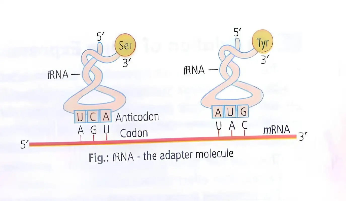
- tRNA reads genetic code and binds specific amino acids; also called soluble RNA (sRNA).
- Secondary structure resembles clover-leaf; actual shape is compact inverted L.
- Has anticodon loop with bases complementary to mRNA codon.
- Has amino acid acceptor end for binding amino acids.
- Each tRNA is specific for one amino acid.
- Initiator tRNA starts translation; no tRNA for stop codons.
Memory Aids
- tRNA = adapter between mRNA codon and amino acid.
- Anticodon pairs with mRNA codon.
- Initiator tRNA starts protein synthesis.
- No tRNA for stop codons.
Quick Checkpoints
- What is the role of tRNA in protein synthesis?
- Describe the structure of tRNA.
- What is an anticodon?
- Which tRNA initiates translation?
- Are there tRNAs for stop codons?
Translation
Process Overview
- Translation is the polymerisation of amino acids into a polypeptide.
- Amino acid sequence is determined by mRNA base sequence.
- Amino acids are joined by peptide bonds, requiring energy.
- Amino acids are activated with ATP and linked to their specific tRNA (charging/aminoacylation).
- Ribosome, made of rRNAs and ~80 proteins, synthesises proteins.
- Ribosome has two subunits: large and small; translation starts when small subunit binds mRNA.
- Large subunit has two sites for amino acid binding; ribosome acts as catalyst (ribozyme) for peptide bond formation.
- mRNA has untranslated regions (UTRs) at 5' and 3' ends for efficient translation.
- Translation starts at AUG codon recognised by initiator tRNA.
- Elongation: aminoacyl-tRNAs bind codons on mRNA; ribosome moves codon to codon adding amino acids.
- Termination: release factor binds stop codon, releasing polypeptide.
Memory Aids
- "Charging tRNA" = amino acid + tRNA + ATP.
- Ribosome = protein factory with two subunits.
- Start codon AUG = initiator tRNA binding site.
- Peptide bond formation catalysed by ribosomal RNA (ribozyme).
- UTRs ensure translation efficiency.
Quick Checkpoints
- What defines amino acid sequence in translation?
- What is the role of tRNA charging?
- How does ribosome catalyse peptide bond formation?
- What are untranslated regions in mRNA?
- How does translation terminate?
Protein Biosynthesis
Post-Translational Modifications
- Proteins made by ribosomes undergo modifications to become mature.
- Modifications are enzyme-catalysed and target specific protein sequences.
Memory Aids
- PTMs = protein maturation steps after synthesis.
- Enzymes recognise specific sequences for modification.
Quick Checkpoints
- What happens to proteins after synthesis?
- What catalyses post-translational modifications?
Polyribosomes
Simultaneous Translation
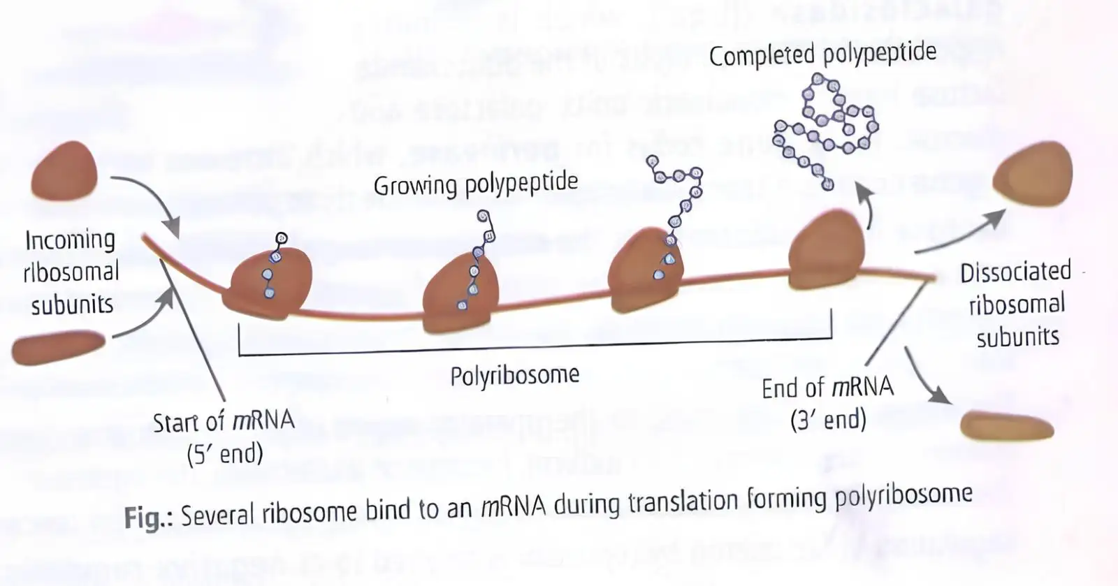
- Multiple ribosomes translate a single mRNA simultaneously.
- First ribosome moves past start codon, allowing next ribosome to bind.
- Cluster of ribosomes on mRNA is called polyribosome.
- Found in both prokaryotes and eukaryotes.
Memory Aids
- Polyribosome = many ribosomes on one mRNA.
- Increases protein production speed.
Quick Checkpoints
- What is a polyribosome?
- How does polyribosome formation increase protein synthesis?
Regulation of Gene Expression
Gene Expression and Regulation
- Genes express to perform specific functions, e.g., beta-galactosidase in E. coli hydrolyses lactose into glucose and galactose.
- Gene expression depends on metabolic, physiological, or environmental conditions.
- Two types of gene regulation: positive (inducible) and negative (repressible).
- Negative regulation: genes express until suppressed by a regulatory product.
- Positive regulation: genes remain off until induced.
- Eukaryotic regulation occurs at transcription, RNA processing, mRNA transport, and translation levels.
- In prokaryotes, gene expression is mainly controlled at transcription initiation.
- RNA polymerase activity at promoters is regulated by proteins binding to operators adjacent to promoters.
- Each operon has a specific operator and repressor, e.g., lac operator binds only lac repressor.
Memory Aids
- Negative = Repressible = Always ON until turned OFF.
- Positive = Inducible = Always OFF until turned ON.
- Prokaryotes control gene expression mainly at transcription initiation.
- Operators are specific to their repressors.
Quick Checkpoints
- What triggers gene expression in positive regulation?
- Where does eukaryotic gene regulation occur?
- What is the role of operator in prokaryotic gene regulation?
- How does negative regulation differ from positive regulation?
The Lac Operon
Structure and Function
- Discovered by Francois Jacob and Jacques Monod.
- Operon: cluster of genes under a single promoter, common in bacteria.
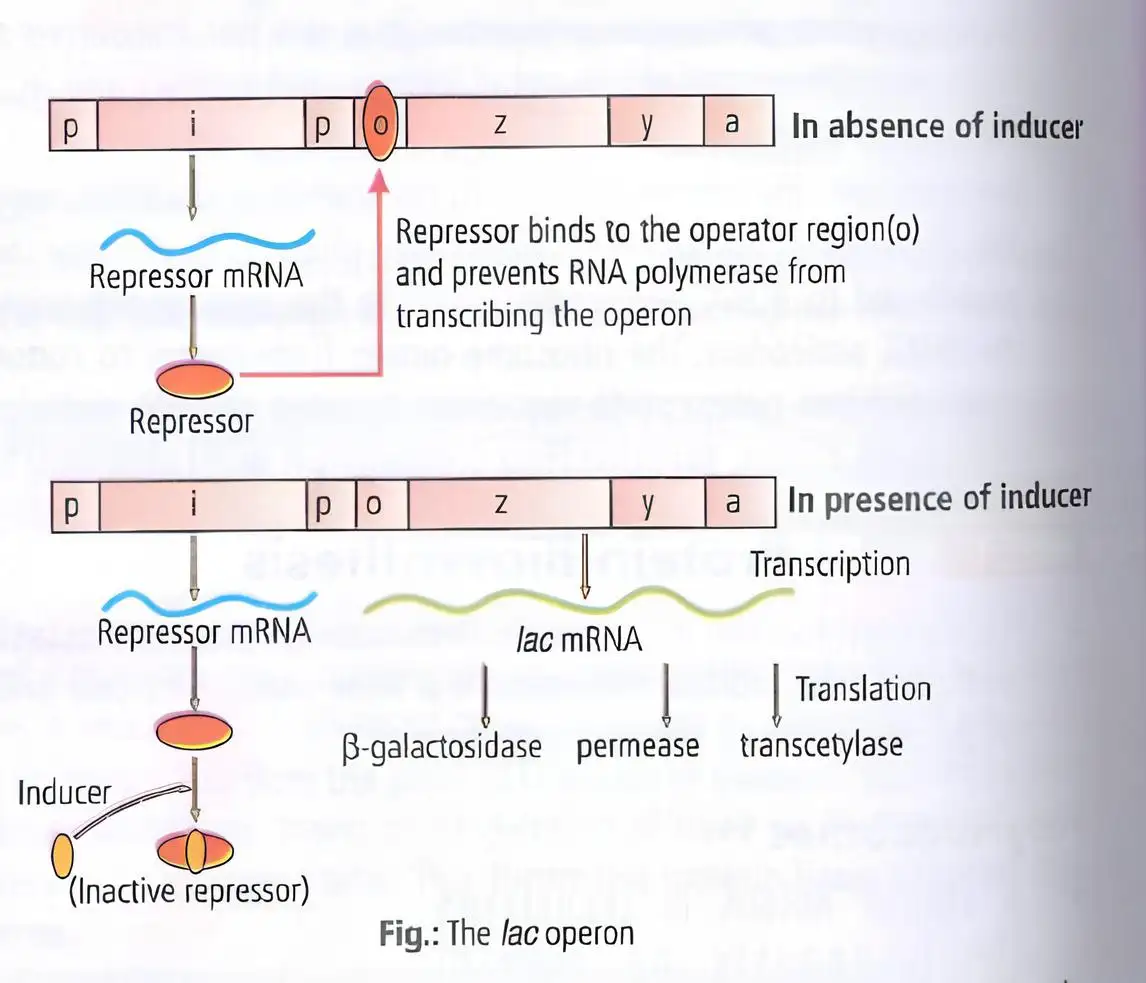
- Lac operon has one regulatory gene (i) and three structural genes (z, y, a).
- i gene codes for lac repressor protein.
- z gene codes for beta-galactosidase, hydrolyzing lactose into glucose and galactose.
- y gene codes for permease, increasing cell permeability to lactose.
- a gene codes for transacetylase, involved in lactose metabolism.
- Lactose acts as an inducer by binding to the repressor and inactivating it.
- In absence of glucose, lactose enters cell via permease and induces operon expression.
- Repressor binds operator to block RNA polymerase and prevent transcription.
- Inducer binding inactivates repressor, allowing transcription of z, y, a genes.
- Regulation by repressor is negative regulation.
Memory Aids
- i gene = inhibitor (repressor).
- z = zero (beta-galactosidase breaks lactose).
- y = yes (permease helps lactose entry).
- a = acetylase (transacetylase enzyme).
- Lactose = inducer that inactivates repressor.
- Negative regulation = repressor blocks transcription until inducer present.
Quick Checkpoints
- What genes are present in lac operon and their functions?
- How does lactose regulate lac operon?
- What is the role of the repressor protein?
- Define negative regulation in lac operon context.
Human Genome Project
Overview
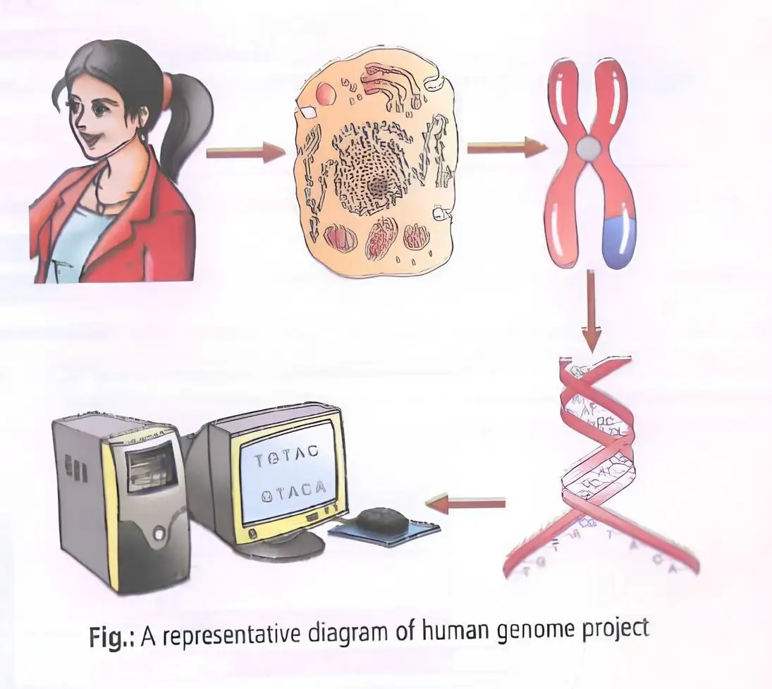
- Launched in 1990 as a mega project called International Human Genome Sequencing Consortium.
- Aimed to identify all human genes, determine their functions, and understand organism formation.
- Coordinated by U.S. Department of Energy and National Institute of Health; completed in 2003.
- Closely linked to bioinformatics development.
Goals
- Identify 20,000-25,000 human genes.
- Sequence 3 billion DNA base pairs.
- Store data in databases.
- Improve data analysis tools.
- Transfer technology to industries.
- Address ethical, legal, social issues (ELSI).
- Develop genetic linkage map using genetic markers.
- Obtain physical map by cloning DNA into BACs and YACs.
Enabling Factors
- Genetic engineering for DNA isolation and cloning.
- Fast DNA sequencing techniques.
- Sequencing of model organisms like bacteria, yeast, Caenorhabditis elegans, Drosophila, rice, and Arabidopsis.
Memory Aids
- HGP = Human genes + sequencing + bioinformatics + ethics.
- Goals: Identify, Sequence, Store, Analyze, Transfer, Ethics, Maps.
Quick Checkpoints
- What is the main aim of HGP?
- Which organizations coordinated HGP?
- What are BACs and YACs used for?
Methodologies of HGP
Techniques Used
- Expressed Sequence Tags (ESTs) to identify genes expressed as RNA.
- Sequence Annotation to assign functions to coding and non-coding regions.
- Automated DNA sequencing based on Sanger's method.
- Generation of overlapping DNA fragments for sequencing.
- Computer programs for sequence alignment and annotation.
- Chromosome 1 sequencing completed in 2006 (last human chromosome sequenced).
Memory Aids
- ESTs focus on expressed genes.
- Sanger method for sequencing.
- Computers essential for sequence assembly.
Quick Checkpoints
- What is the role of ESTs?
- Why are computers needed in HGP?
- When was chromosome 1 sequencing completed?
Salient Features of HGP
Key Observations
- Human genome has 3,164.7 million nucleotide base pairs.
- Average gene size ~3000 bases; largest gene dystrophin (2.4 million bases).
- Estimated 30,000 genes; 99.9% nucleotide bases identical in all humans.
- Functions of over 50% genes unknown.
- Less than 2% genome codes for proteins.
- Repeated DNA sequences form large genome portion.
- Chromosome 1 has most genes (2968); Y chromosome has fewest (231).
- About 1.4 million single nucleotide polymorphisms (SNPs) identified.
Memory Aids
- Genome size ~3.2 billion bases.
- Most genes on chromosome 1, least on Y.
- SNPs = single base differences in DNA.
Quick Checkpoints
- How many base pairs are in the human genome?
- What percentage of genome codes for proteins?
- What are SNPs?
Applications of HGP
Uses
- Enables systematic, large-scale biological research.
- Allows study of all genes or transcripts in tissues/organs/tumors.
- Helps understand gene and protein networks in life chemistry.
Memory Aids
- HGP = Research scale-up + gene study + network understanding.
Quick Checkpoints
- How does HGP change biological research?
- What can be studied using HGP data?
- Why is understanding gene networks important?
DNA Fingerprinting
Concept and Process
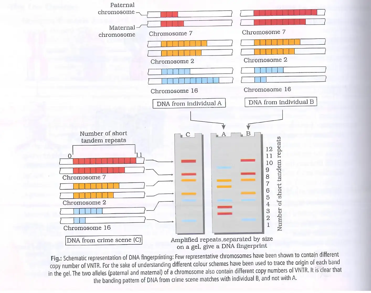
- DNA fingerprinting identifies differences in repetitive DNA regions called Variable Number of Tandem Repeats (VNTRs).
- VNTR size ranges from 0.1 to 20 kb; repetitive DNA forms satellite DNA peaks separate from bulk genomic DNA.
- These sequences show high polymorphism, unique to individuals except identical twins.
- DNA from any tissue (blood, hair, skin, saliva, sperm) shows the same polymorphism pattern.
- Useful in forensic identification and genetic diversity studies.
- Polymorphism arises from mutations; multiple alleles exist at a locus with frequency >0.01.
- Developed by Alec Jeffreys using satellite DNA probes and Southern blot hybridization.
- Steps: DNA extraction, PCR amplification, restriction enzyme digestion, electrophoresis, denaturation, transfer to membrane (Southern blotting), probe hybridization, autoradiography.
Memory Aids
- VNTR = Variable Number of Tandem Repeats, key to uniqueness.
- Polymorphism = multiple alleles due to mutations.
- Southern blotting = transfer DNA to membrane for probe detection.
- Alec Jeffreys = pioneer of DNA fingerprinting.
Quick Checkpoints
- What is VNTR and its role in DNA fingerprinting?
- How does polymorphism contribute to individual identification?
- What are the main steps in DNA fingerprinting?
- Who developed DNA fingerprinting and what probe was used?
Southern Blotting
Technique and Steps
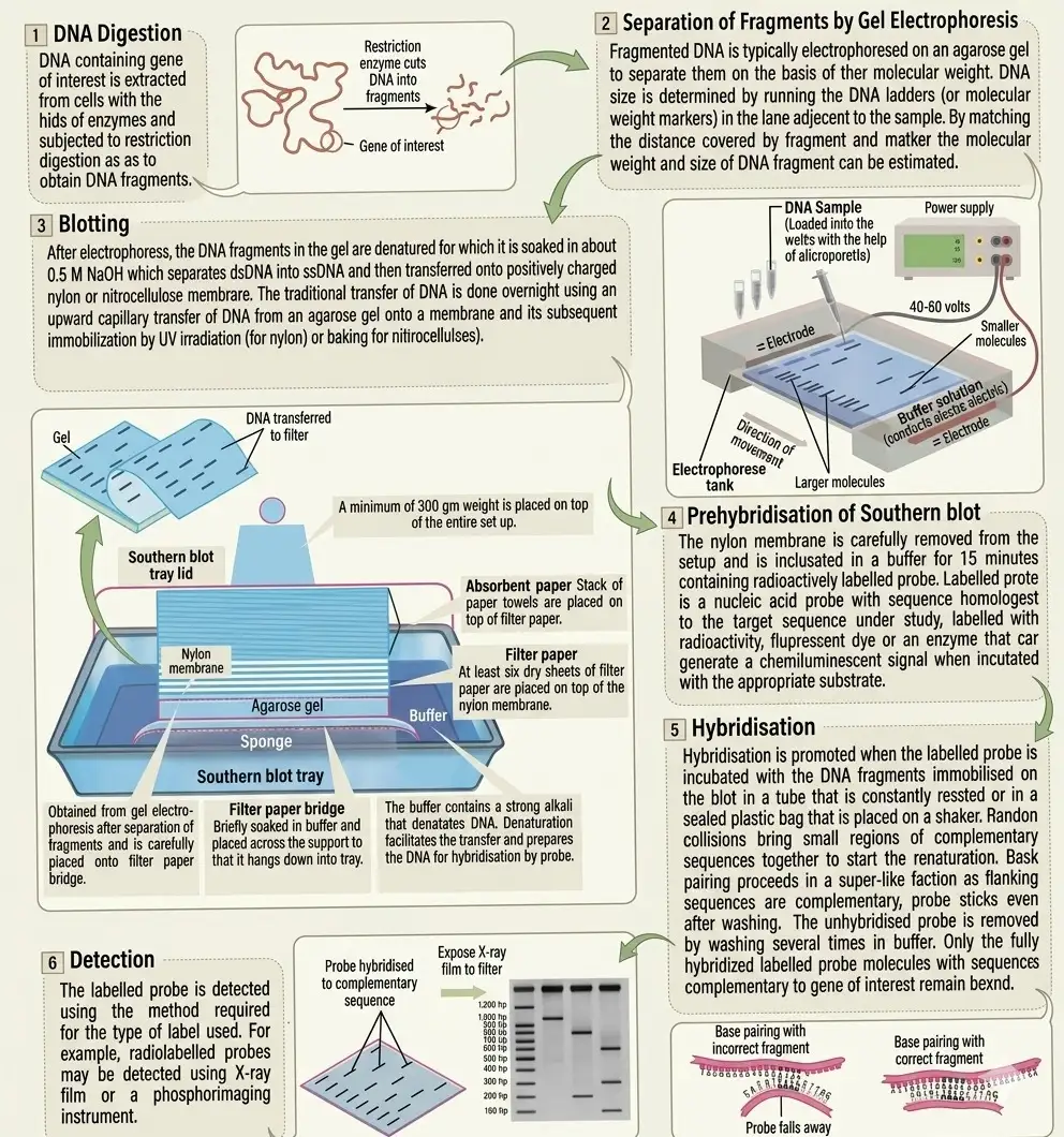
- Transfers DNA fragments from gel to membrane, preserving banding pattern.
- DNA immobilized on membrane for hybridization with labeled probes.
- Named after Edwin Southern (1975).
- Steps: DNA digestion by restriction enzymes, gel electrophoresis, transfer to nylon membrane, prehybridization with buffer, hybridization with specific probe, detection by X-ray or other methods.
- Used to detect specific DNA sequences within a mixture.
Memory Aids
- Southern blot = DNA transfer and detection.
- Restriction enzymes cut DNA into fragments.
- Probe binds only to complementary DNA sequences.
- Detection shows dark bands where probe hybridizes.
Quick Checkpoints
- What is the purpose of Southern blotting?
- How are DNA fragments separated before transfer?
- What role does the probe play in Southern blotting?
- How is the hybridized DNA detected?
DNA Sequencing by Sanger's Method
Process and Significance
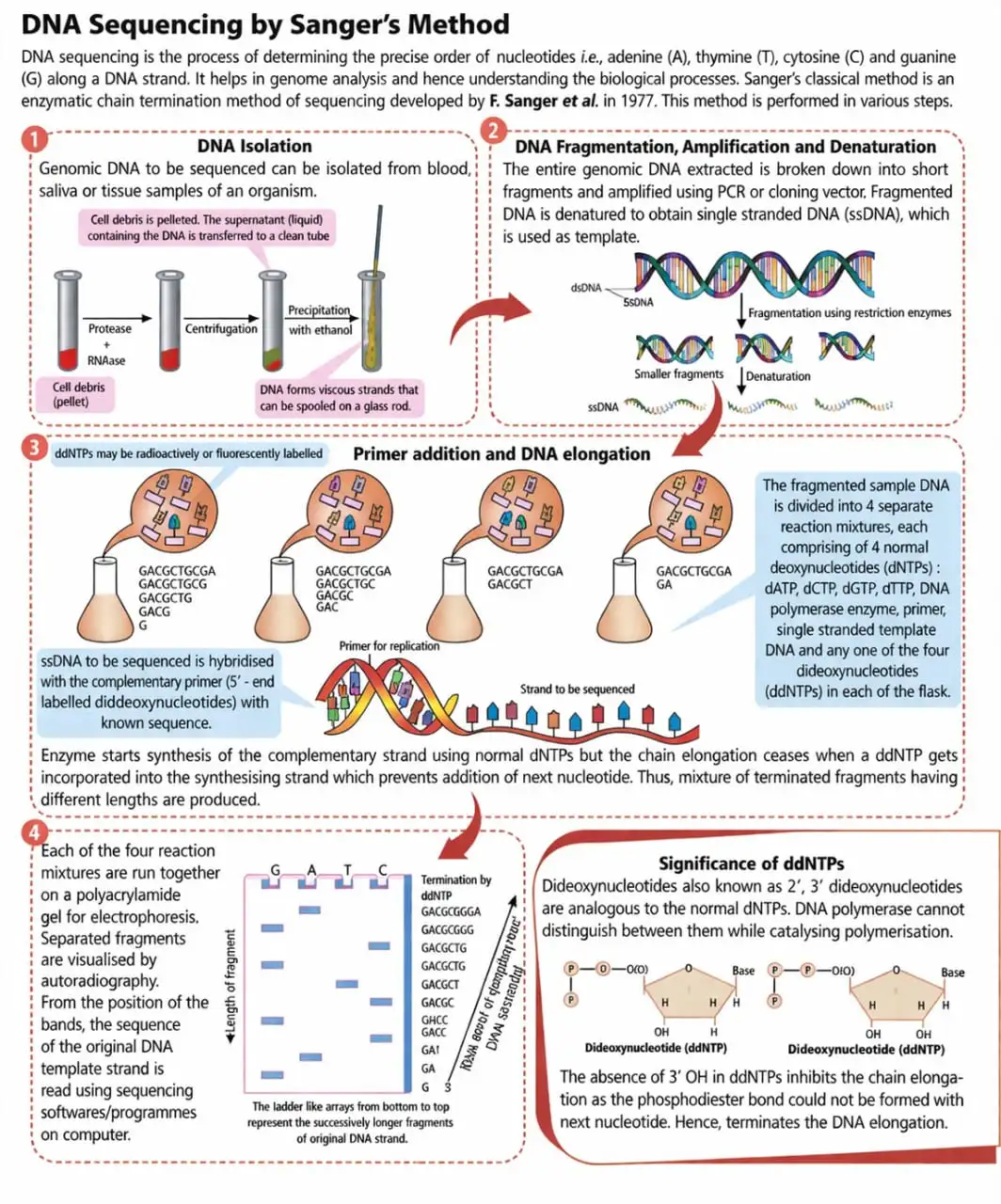
- Determines exact nucleotide order (A, T, C, G) in DNA.
- Developed by F. Sanger in 1977 using enzymatic chain termination.
- Steps: DNA isolation, fragmentation, PCR amplification, denaturation, primer binding, DNA polymerase elongation with normal nucleotides and chain-terminating ddNTPs.
- ddNTPs lack 3' OH group, causing chain termination at random points.
- Fragments separated by gel electrophoresis; sequence deduced from fragment sizes.
- Essential for genome analysis and understanding biological processes.
Memory Aids
- ddNTPs stop DNA chain growth, enabling sequence determination.
- Chain termination method = Sanger sequencing.
- Gel electrophoresis separates fragments by size.
- Primer initiates DNA synthesis.
Quick Checkpoints
- What causes chain termination in Sanger sequencing?
- Why is gel electrophoresis used in sequencing?
- What is the role of primers in DNA sequencing?
- How does Sanger sequencing help in genome analysis?
NEET Botany — ALL CHAPTERS
1
The Living World
2
Plant Kingdom
3
Morphology of Flowering Plants
4
Anatomy of Flowering Plants
5
The Unit of Life
6
Photosynthesis in Higher Plants
7
Respiration in Plants
8
Plant Growth and Development
9
Sexual Reproduction in Flowering Plants
10
Principles of Inheritance and Variation
11
Molecular Basis of Inheritance
12
Biotechnology :Principles and Processes
13
Biotechnology and its Applications