NEET Ug . NEET Botany . Botany . Anatomy of Flowering Plants
Chapter 4: Anatomy of Flowering Plants
Ch 4
NEET Botany
NEET Ug
Introduction
Overview
- Plant anatomy is the study of internal plant structure.
- Plants have cells organized into tissues, which form organs.
- Monocots and dicots differ anatomically within angiosperms.
- Katherine Esau showed that curly top virus spreads via phloem.
- She authored key books: "Plant Anatomy" (1954) and "The Anatomy of Seed Plants" (1960).
Memory Aids
- Esau = Phloem virus spread + Plant anatomy books.
- Cells → Tissues → Organs hierarchy.
Quick Checkpoints
- What is plant anatomy?
- How do monocots and dicots differ anatomically?
- Which tissue spreads the curly top virus?
The Tissues
Classification
- Tissue: group of cells with common origin and function.
- Two main types: meristematic (dividing) and permanent (non-dividing).
- Term coined by N. Grew in 1682.
Memory Aids
- Meristematic = dividing cells.
- Permanent = mature, non-dividing cells.
Quick Checkpoints
- Define tissue in plants.
- What are the two main tissue types?
Meristematic Tissues
Characteristics
- Regions of active cell division called meristems (meristos = divide).
- Cells are immature, isodiametric or polygonal, with thin, flexible walls.
- No intercellular spaces; cells compactly arranged.
- Vacuoles absent or small; large nucleus and dense cytoplasm.
- Term coined by C. Nageli in 1858.
Memory Aids
- Meristem = dividing, immature cells with thin walls.
- No vacuoles or intercellular spaces.
Quick Checkpoints
- What defines meristematic tissue?
- Describe meristematic cell features.
Classification of Meristem
Types by Origin and Position
- Based on origin: promeristem, primary meristem, secondary meristem.
- Promeristem: embryonic meristem forming the plant embryo.
- Primary meristem: develops from promeristem; forms primary plant body; found at apices.
- Secondary meristem: arises by dedifferentiation from primary permanent tissue; forms secondary plant body.
- Based on position: apical (tips), intercalary (between mature tissues), lateral (cylindrical, in mature regions).
Memory Aids
- Promeristem = embryo stage.
- Primary = apical, early growth.
- Secondary = dedifferentiated, secondary growth.
- Apical = tips; Intercalary = between tissues; Lateral = cylindrical.
Quick Checkpoints
- What are the three meristem types by origin?
- Where are apical meristems located?
- What is the role of secondary meristem?
Apical Meristem
Features
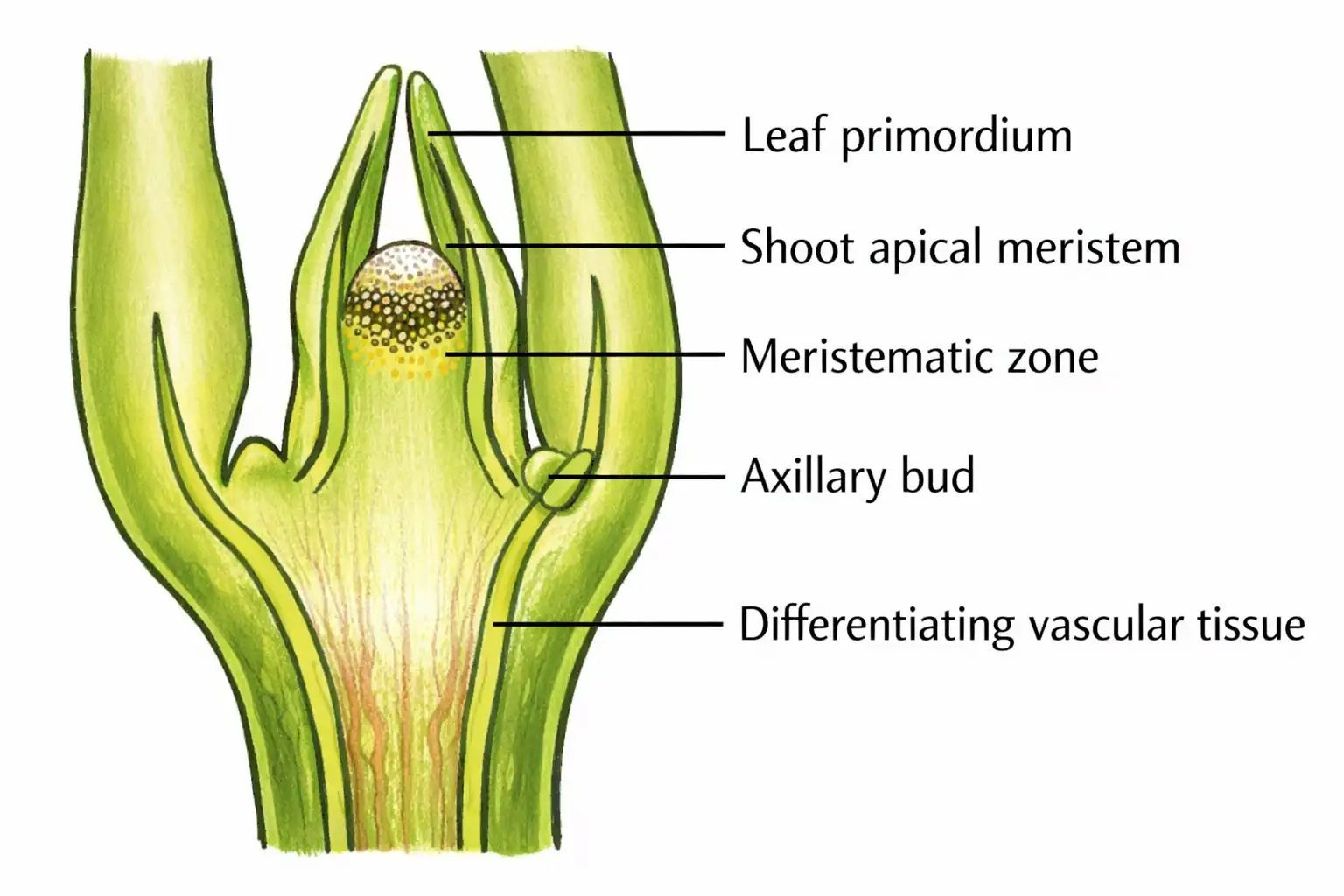
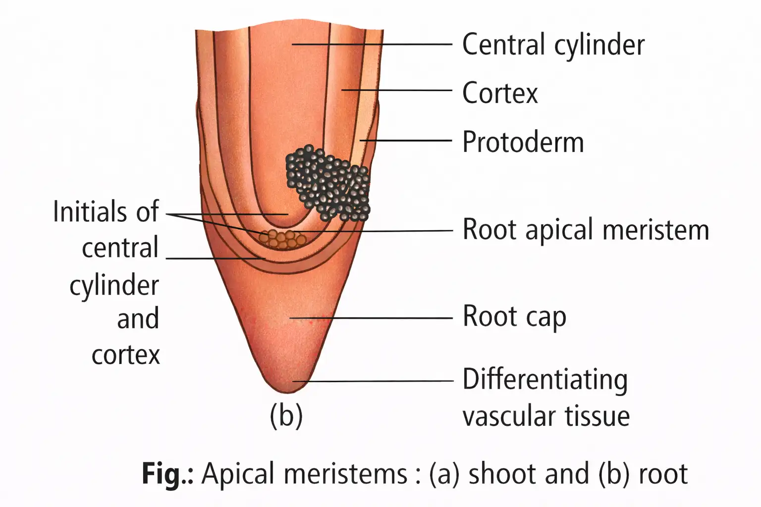
- Located at root and shoot tips; produces primary tissues.
- Root apical meristem at root tip; shoot apical meristem at stem apex.
- Cells left behind from shoot apical meristem form axillary buds (can form branches or flowers).
Memory Aids
- Apical = apex (tips) growth.
- Axillary buds = leftover cells from shoot apex.
Quick Checkpoints
- Where is the shoot apical meristem located?
- What forms axillary buds?
Intercalary Meristem
Features
- Located between mature tissues, common in grasses.
- Enables regeneration after grazing.
- Both apical and intercalary meristems are primary meristems.
Memory Aids
- Intercalary = between mature tissues.
- Found in grasses for regrowth.
Quick Checkpoints
- Where is intercalary meristem found?
- Why is it important in grasses?
Lateral Meristem
Features
- Occurs in mature regions of roots and shoots, especially woody plants.
- Also called secondary meristem; cylindrical in shape.
- Examples: fascicular vascular cambium, interfascicular cambium, cork cambium.
- Responsible for secondary growth (thickening).
- Vegetative shoot apex is terminal meristem at stem tips; root apex is sub-terminal, protected by root cap.
- Quiescent centre: group of distal root cells with low mitotic activity, acting as a reserve.
Memory Aids
- Lateral = secondary growth, cylindrical cambium.
- Quiescent centre = root cell reserve.
Quick Checkpoints
- What is the function of lateral meristem?
- Where is the quiescent centre located?
Permanent Tissues
Features
- Cells lose division ability, become specialized.
- Two types: simple (all cells similar) and complex (different cell types).
Memory Aids
- Permanent = mature, non-dividing cells.
- Simple = uniform cells; Complex = mixed cells.
Quick Checkpoints
- Define permanent tissue.
- Difference between simple and complex tissues?
Simple Tissues
Types
- Made of one cell type: parenchyma, collenchyma, sclerenchyma.
Memory Aids
- Simple = one cell type.
Quick Checkpoints
- Name the simple tissues in plants.
Parenchyma
Features
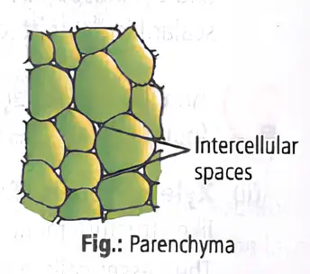
- Major component of organs; cells isodiametric, spherical, oval, polygonal or elongated.
- Thin cellulose walls; may have small intercellular spaces.
- Functions: photosynthesis, storage, secretion.
- Chlorenchyma: parenchyma with chloroplasts in leaf mesophyll for photosynthesis.
- Aerenchyma: parenchyma with large air spaces in aquatic plants for buoyancy.
Memory Aids
- Parenchyma = basic tissue, thin walls, various shapes.
- Chlorenchyma = photosynthetic parenchyma.
- Aerenchyma = air spaces for buoyancy.
Quick Checkpoints
- What are the functions of parenchyma?
- Define chlorenchyma and aerenchyma.
Collenchyma
Features
- Supporting tissue below epidermis in dicots; in layers or patches.
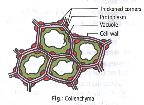
- Cells thickened at corners by cellulose, hemicellulose, pectin.
- Cells oval, spherical or polygonal; may contain chloroplasts.
- No intercellular spaces; provides mechanical support to young stems and petioles.
Memory Aids
- Collenchyma = unevenly thickened corners, flexible support.
Quick Checkpoints
- Where is collenchyma found?
- What is the function of collenchyma?
Sclerenchyma
Features
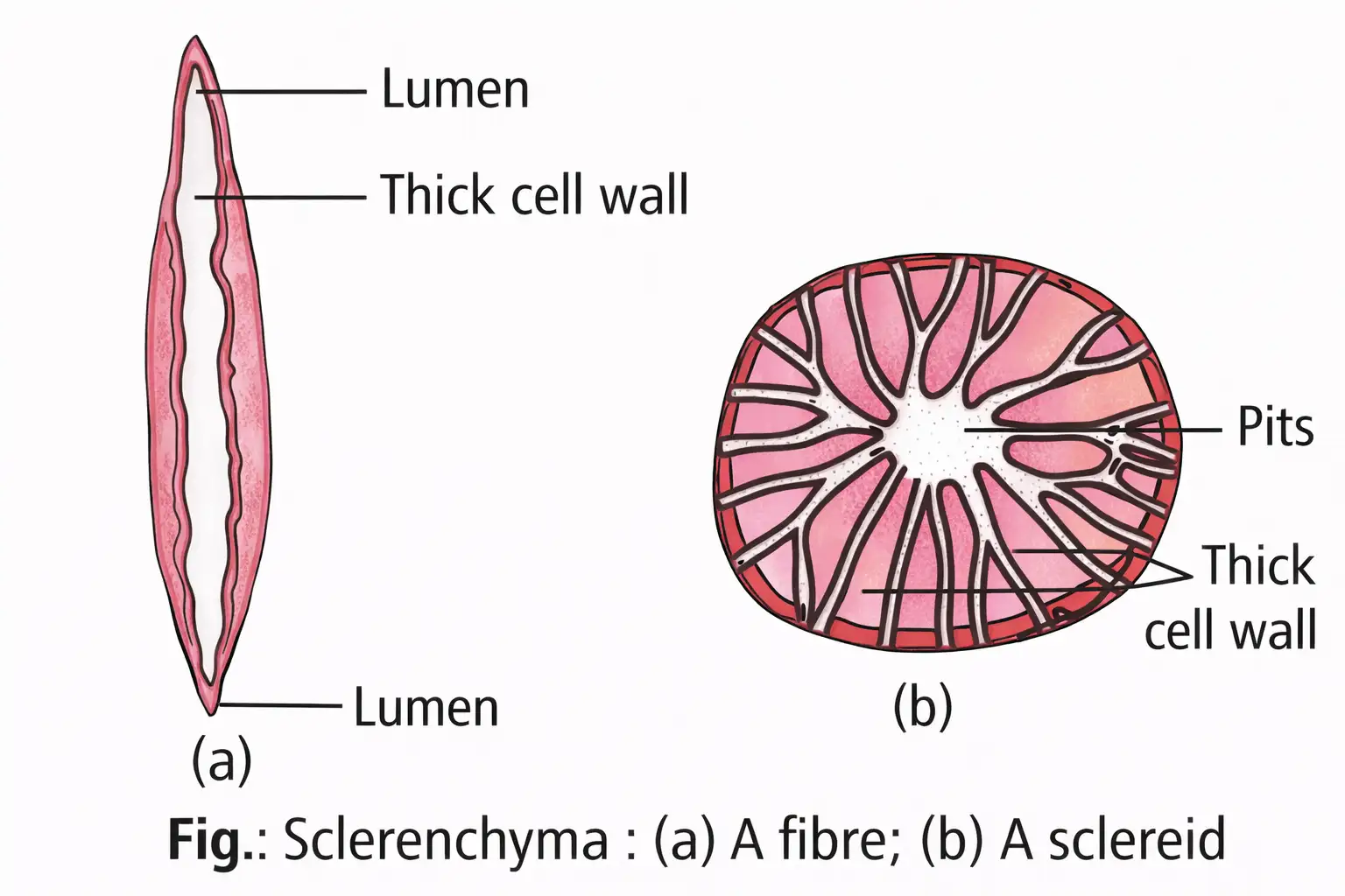
- Cells with thick lignified walls; provide mechanical support.
- Usually dead at maturity; walls have pits.
- Two types: fibres (elongated, pointed ends, with lumen and pits) and sclereids (short, thick-walled, narrow lumen).
- Found in fruit walls, seed coats, leaves, and coir (coconut fibre).
Memory Aids
- Sclerenchyma = thick lignified walls, dead cells.
- Fibres = long; Sclereids = short, irregular.
Quick Checkpoints
- What are the two types of sclerenchyma?
- Where is sclerenchyma commonly found?
Complex Tissues
Features
- Made of different cell types working as a unit.
- Also called vascular or conducting tissues.
- Two types: xylem and phloem.
Memory Aids
- Complex = multiple cell types, vascular tissues.
Quick Checkpoints
- What are complex tissues?
- Name the two complex tissues.
Xylem
Components and Functions
- Conducts water and minerals from roots to shoots.
- Provides mechanical strength.
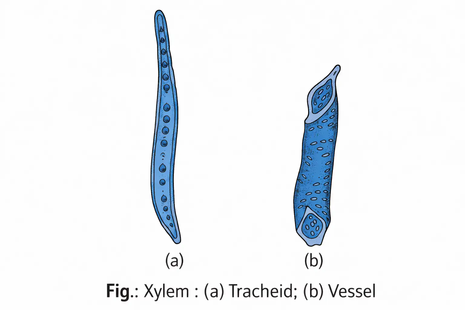
- Composed of tracheids, vessels, xylem fibres, and xylem parenchyma.
- Tracheids: elongated, lignified, dead cells with tapered ends and pits; primitive water conductors.
- Vessels: long tubes of vessel members with perforated end walls; characteristic of angiosperms.
- Xylem fibres: sclerenchyma fibres with thick walls and obliterated lumen.
- Xylem parenchyma: living cells storing food and aiding radial water conduction.
- Primary xylem: protoxylem (first formed) and metaxylem (later formed).
- Arrangement: endarch in stems (protoxylem inside), exarch in roots (protoxylem outside).
- Secondary xylem: formed during secondary growth; no protoxylem/metaxylem differentiation.
Memory Aids
- Xylem = water conduction + support.
- Tracheids = primitive; Vessels = advanced.
- Protoxylem inside stem, outside root.
Quick Checkpoints
- What are the four elements of xylem?
- Difference between protoxylem and metaxylem?
- What is endarch and exarch arrangement?
Phloem
Components and Functions
- Transports food from leaves to other parts.
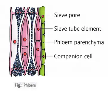
- In angiosperms: sieve tube elements, companion cells, phloem parenchyma, phloem fibres.
- In gymnosperms: sieve cells and albuminous cells; lack sieve tubes and companion cells.
- Sieve tube elements: long tubes with sieve plates; lack nucleus; controlled by companion cells.
- Companion cells: specialized parenchyma; maintain sieve tube function; connected by pit fields.
- Phloem parenchyma: elongated cells with pits; store food and substances; absent in most monocots.
- Phloem fibres: thick-walled, dead at maturity; called bast fibres; commercial fibers from jute, hemp, flax.
- Primary phloem: protophloem (first formed, narrow sieve tubes) and metaphloem (later formed, wider sieve tubes).
- Special cells in pteridophytes and gymnosperms: albuminous or Strasburger cells.
Memory Aids
- Phloem = food transport.
- Sieve tubes + companion cells = functional unit.
- Bast fibres = commercial fibers.
Quick Checkpoints
- What are the components of phloem in angiosperms?
- Difference between sieve tube elements and companion cells?
- What are bast fibres?
Epidermal Tissue System
Epidermis
- Single-layered outermost covering of primary plant body.
- Composed of parenchymatous cells with large vacuole and thin cytoplasm.
- Outer surface covered by waxy cuticle to prevent water loss; absent in roots.
Stomata
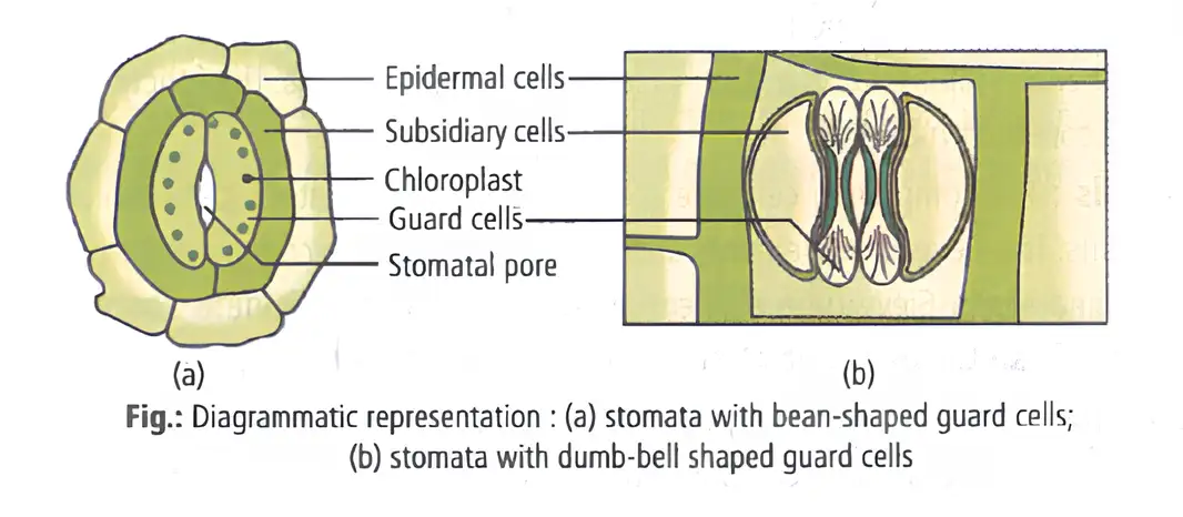
- Present in leaf epidermis; regulate transpiration and gas exchange.
- Each stoma has two guard cells (bean-shaped or dumbbell-shaped in grasses).
- Guard cells have thin outer walls and thick inner walls; contain chloroplasts.
- Opening and closing of stomata controlled by guard cells.
- Stomatal apparatus = stomatal aperture + guard cells + subsidiary cells.
Epidermal Appendages
- Root hairs: unicellular extensions aiding water and mineral absorption.
- Trichomes: multicellular hairs on stems; may be branched/unbranched, soft/stiff, or secretory.
- Trichomes reduce water loss by transpiration.
Memory Aids
- Epidermis = single layer + cuticle (except roots).
- Stomata = guard cells + subsidiary cells = stomatal apparatus.
- Root hairs absorb; trichomes protect.
Quick Checkpoints
- What is the function of the cuticle?
- Describe the structure of guard cells.
- What forms the stomatal apparatus?
- Difference between root hairs and trichomes?
Ground Tissue System
Components and Structure
- Largest tissue system in plants.
- Includes hypodermis, cortex, endodermis, pericycle, medullary rays, and pith.
- All tissues except epidermis and vascular bundles.
- Made of simple tissues: parenchyma, collenchyma, sclerenchyma.
- In leaves, ground tissue = mesophyll (chloroplast-rich cells).
Memory Aids
- Ground tissue = all non-epidermal, non-vascular tissues.
- Mesophyll = leaf ground tissue with chloroplasts.
Quick Checkpoints
- Which tissues form the ground tissue system?
- What is mesophyll?
- What simple tissues are found in ground tissue?
Vascular Tissue System
Structure and Types
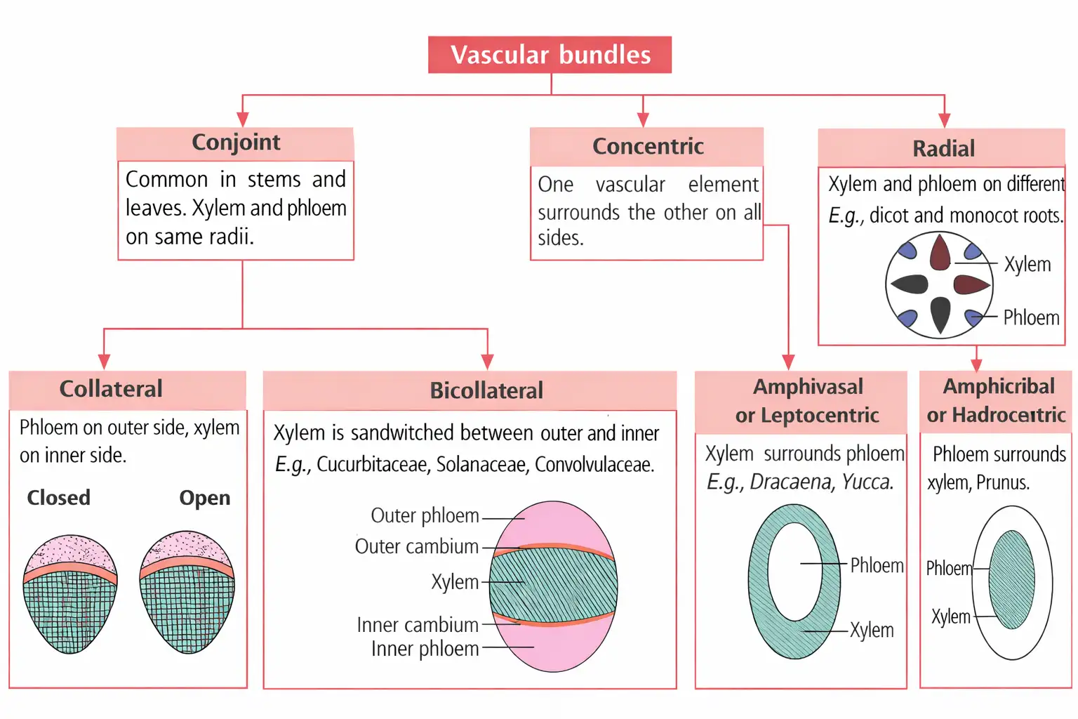
- Continuous tissues from root to leaf called vascular bundles.
- Complex tissues: xylem (water transport) and phloem (food transport).
- Dicot stems have cambium between xylem and phloem; called open vascular bundles (secondary growth possible).
- Monocot vascular bundles lack cambium; called closed vascular bundles (no secondary growth).
- Vascular bundles classified by xylem-phloem arrangement:
- Radial: xylem and phloem on different radii (roots).
- Conjoint: xylem and phloem together on same radius (stems, leaves).
- Collateral: phloem outside, xylem inside; open or closed.
- Bicollateral: xylem between two phloem layers with cambium.
- Concentric: one tissue surrounds the other.
- Amphivasal: xylem surrounds phloem.
- Amphicribral: phloem surrounds xylem.
Memory Aids
- Open bundles = cambium present; closed bundles = no cambium.
- Radial = separate xylem and phloem; conjoint = together; concentric = one surrounds other.
Quick Checkpoints
- What defines open and closed vascular bundles?
- List types of vascular bundles by arrangement.
- Where are radial bundles found?
NEET Botany — ALL CHAPTERS
1
The Living World
2
Plant Kingdom
3
Morphology of Flowering Plants
4
Anatomy of Flowering Plants
5
The Unit of Life
6
Photosynthesis in Higher Plants
7
Respiration in Plants
8
Plant Growth and Development
9
Sexual Reproduction in Flowering Plants
10
Principles of Inheritance and Variation
11
Molecular Basis of Inheritance
12
Biotechnology :Principles and Processes
13
Biotechnology and its Applications