NEET Ug . NEET Zoology . Zoology . Structural Organisation in Animals
Chapter 1: Structural Organisation in Animals
Ch 1
NEET Zoology
NEET Ug
Introduction
Overview
- Unicellular organisms perform all life functions in a single cell.
- Multicellular organisms have specialized groups of cells performing specific functions.
- A tissue is a group of similar cells with common origin and function.
Memory Aids
- Tissue = group of similar cells + intercellular substance + common function.
Quick Checkpoints
- What defines a tissue in multicellular organisms?
- How do unicellular and multicellular organisms differ in function execution?
Animal Tissues
Types and Classification
- Four basic tissue types: Epithelial, Connective, Muscular, Neural.
- Each tissue type has distinct origin and function.
- Epithelial: protection, secretion, absorption, excretion, reproduction.
- Connective: support, attachment, storage, protection, transport.
- Muscular: movement and locomotion.
- Neural: control and nerve impulse transmission.

Memory Aids
- Remember: Epithelial = covering; Connective = support; Muscular = movement; Neural = control.
Quick Checkpoints
- List the four basic animal tissues.
- What are the main functions of connective tissue?
Epithelial Tissue
Structure and Types
- Epithelium has a free surface facing body fluid or environment.
- Cells are tightly packed with minimal intercellular matrix.
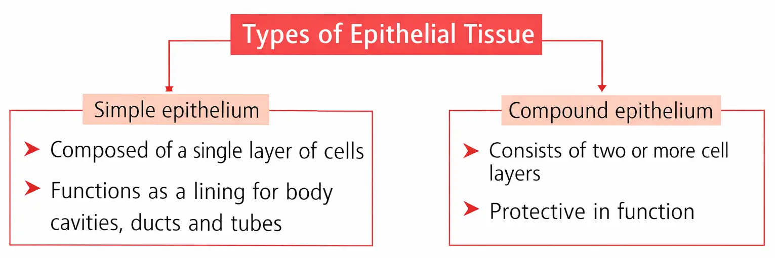
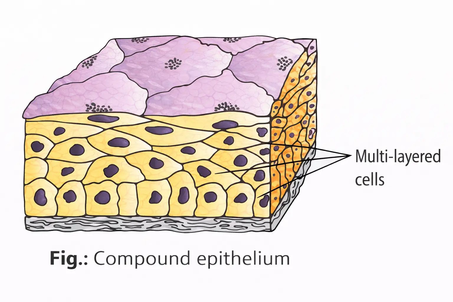
- Two main types: Simple (single layer) and Compound (multiple layers).
- Simple epithelium subtypes: Squamous, Cuboidal, Columnar.
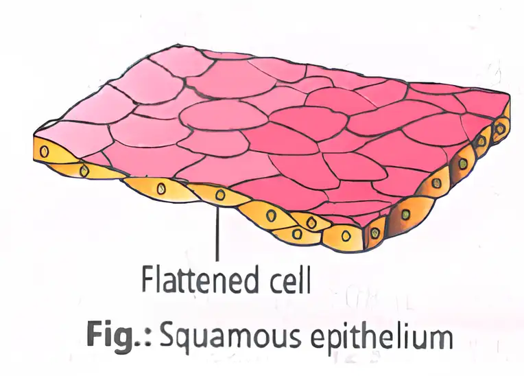
- Squamous: thin, flat cells; found in blood vessels and lungs; functions include protection and gas exchange.
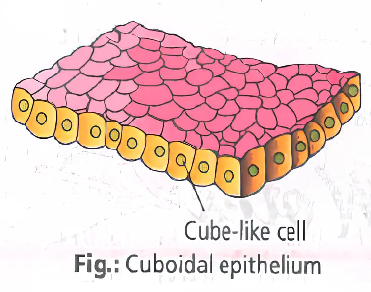
- Cuboidal: cube-shaped cells; found in gland ducts and kidney tubules; functions include secretion and absorption; may have microvilli.
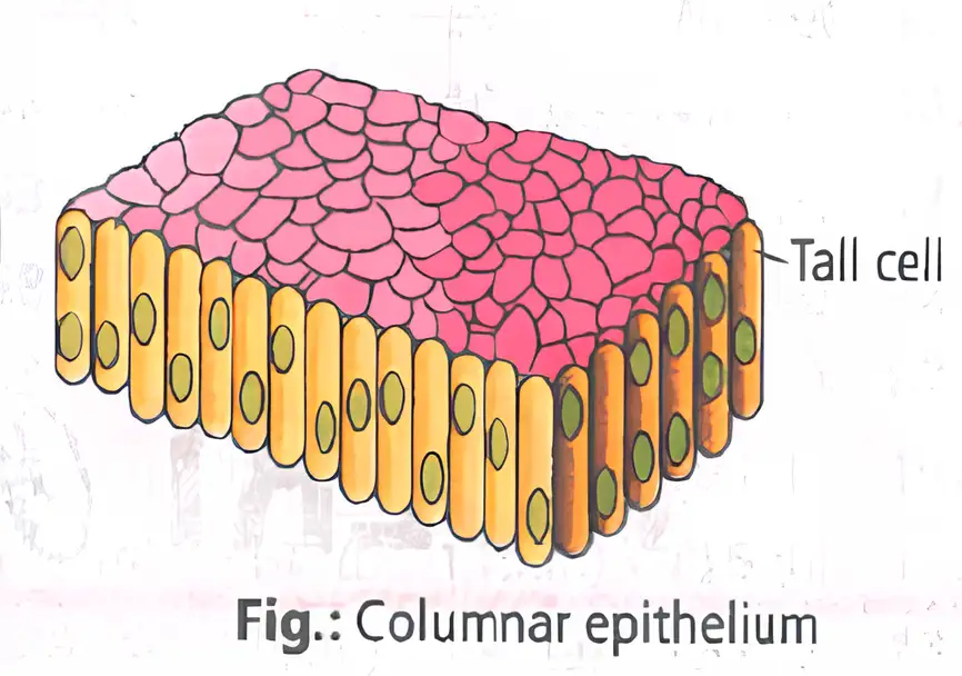
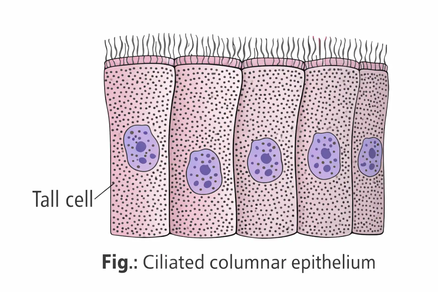
- Columnar: tall cells with basal nuclei; found in stomach, intestine; may have microvilli (brush border) or cilia.
- Ciliated epithelium moves mucus, urine, eggs, cerebrospinal fluid.
- Compound epithelium protects against mechanical and chemical stress; found in skin, mouth lining, ducts.
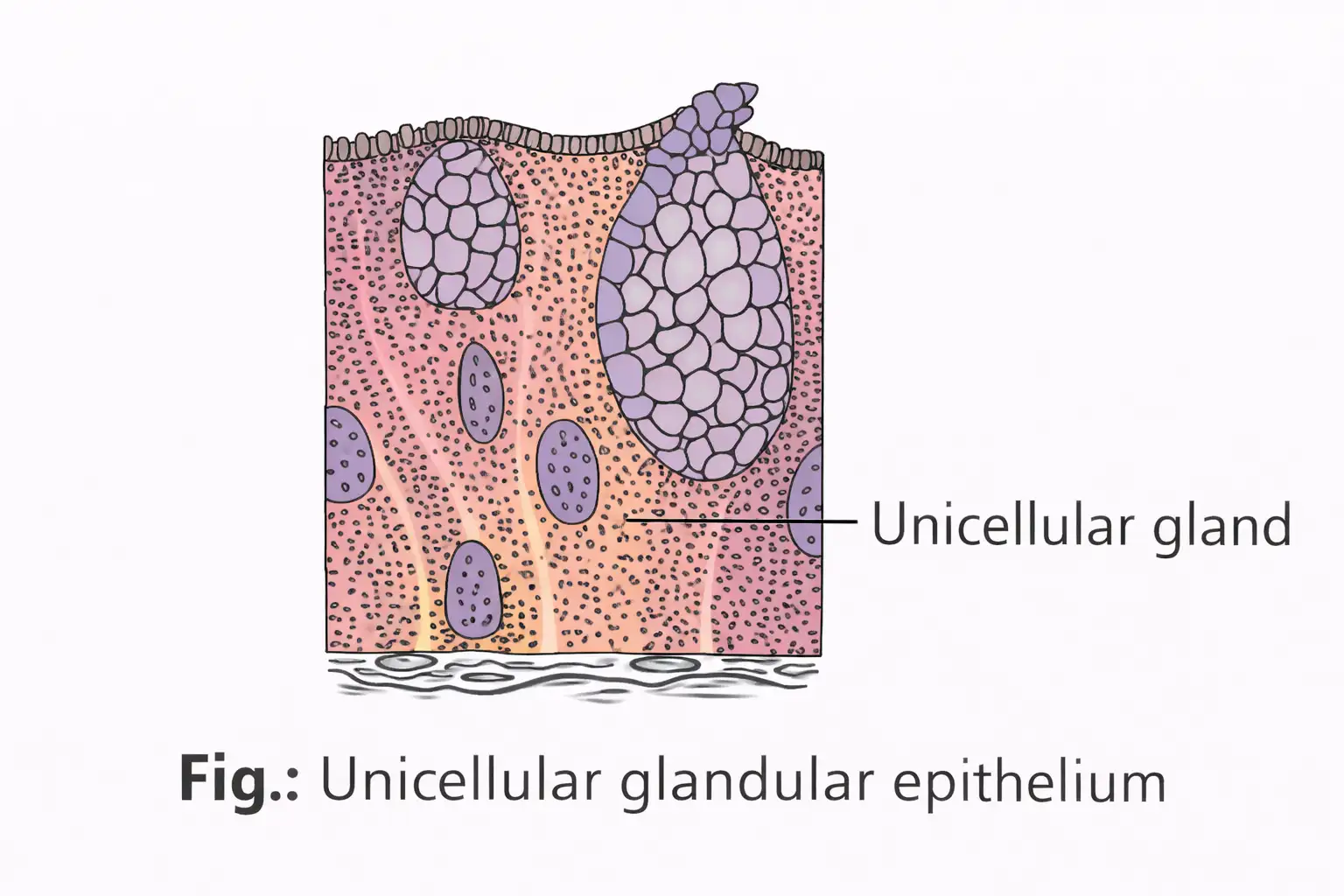
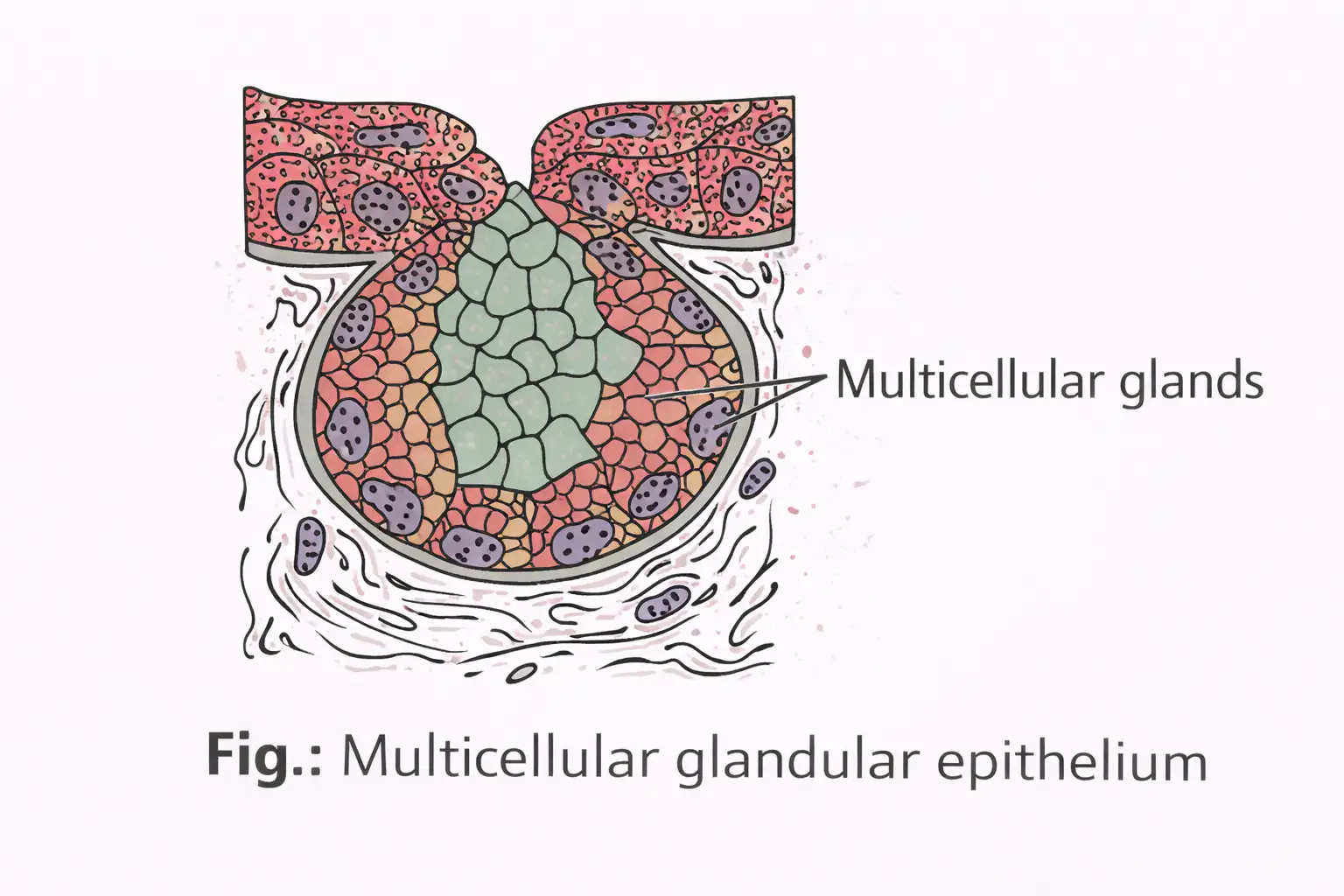
- Glandular epithelium specialized for secretion; unicellular (goblet cells) or multicellular (salivary glands).
- Glands classified as exocrine (ducts) or endocrine (ductless, secrete hormones).
- Heterocrine glands have both exocrine and endocrine parts (e.g., pancreas).
- Cell junctions: Tight (prevent leakage), Adhering (cement cells), Gap (communication).
Memory Aids
- Simple epithelium = 1 layer; Compound = multiple layers.
- Squamous = flat; Cuboidal = cube; Columnar = tall.
- Ciliated epithelium moves substances; glandular epithelium secretes.
- Exocrine glands use ducts; endocrine glands are ductless.
Quick Checkpoints
- What are the types of simple epithelium?
- Where is ciliated epithelium found and what is its function?
- Differentiate exocrine and endocrine glands.
- What are the three types of cell junctions in epithelium?
Connective Tissue
Overview and Types
- Most abundant tissue; links and supports other tissues/organs.
- Cells secrete collagen or elastin fibers for strength and flexibility.
- Matrix contains modified polysaccharides.
- Types: Loose (areolar, adipose), Dense (regular, irregular), Specialized (cartilage, bone, blood).
- Loose connective tissue: cells and fibers loosely arranged in semi-fluid ground substance.
- Dense connective tissue: fibers and fibroblasts densely packed.
- Dense regular: collagen fibers in parallel; found in tendons and ligaments.
- Dense irregular: collagen fibers in multiple directions; found in skin.
- White fibrous connective tissue: collagen bundles, strong but less flexible.
- Yellow elastic connective tissue: elastic fibers, flexible; forms ligaments and vessel walls.
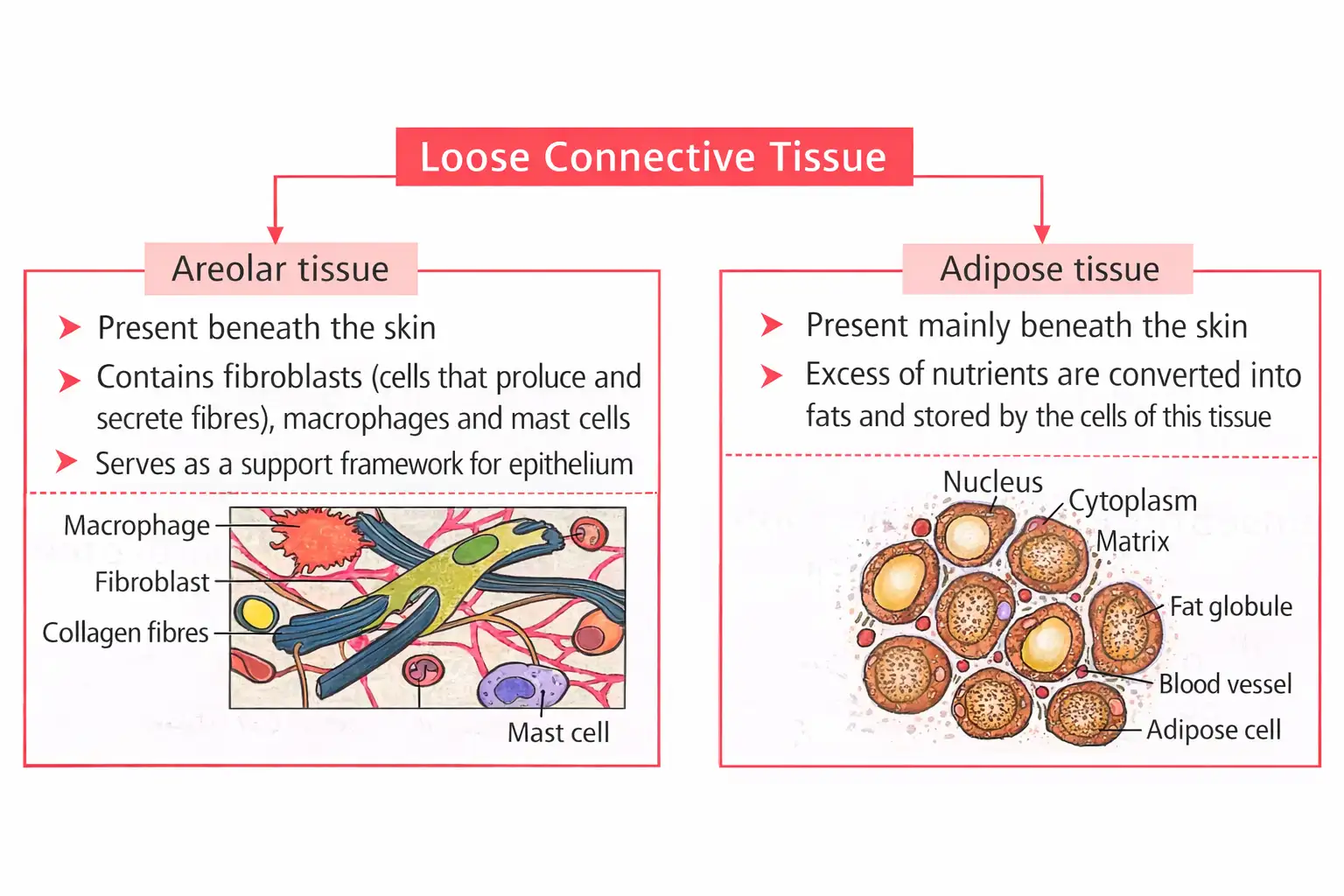
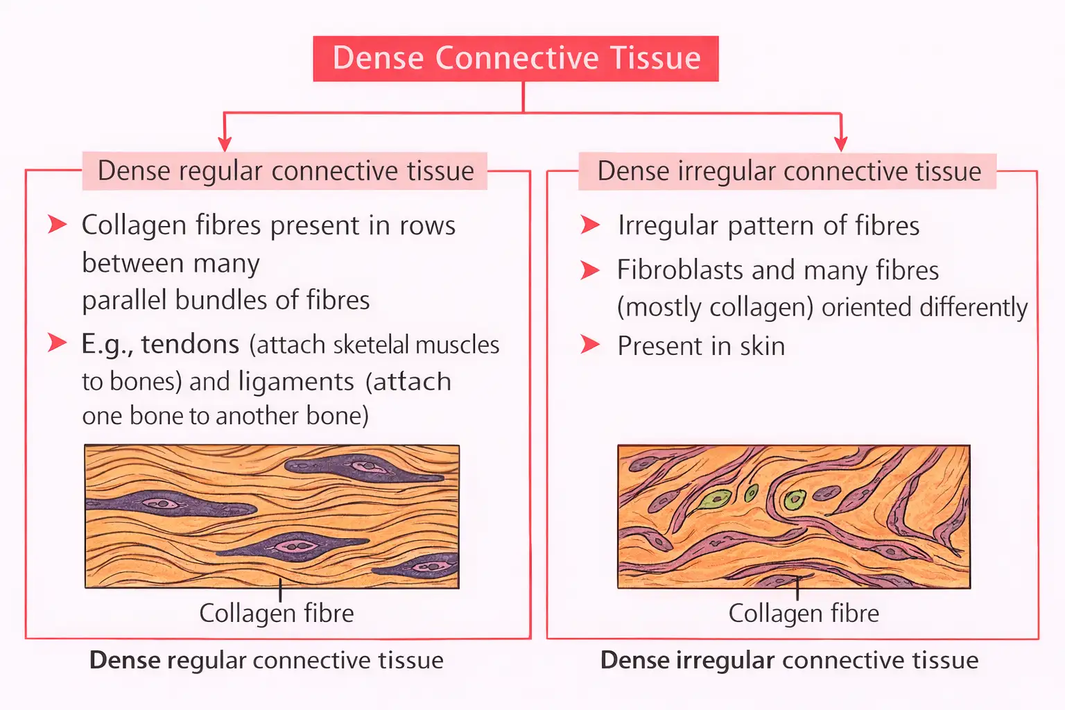
Memory Aids
- Loose = loose fibers; Dense = tightly packed fibers.
- White fibrous = strength; Yellow elastic = elasticity.
Quick Checkpoints
- What are the main functions of connective tissue?
- Differentiate loose and dense connective tissue.
- Where is white fibrous connective tissue found?
Specialised Connective Tissue
Cartilage
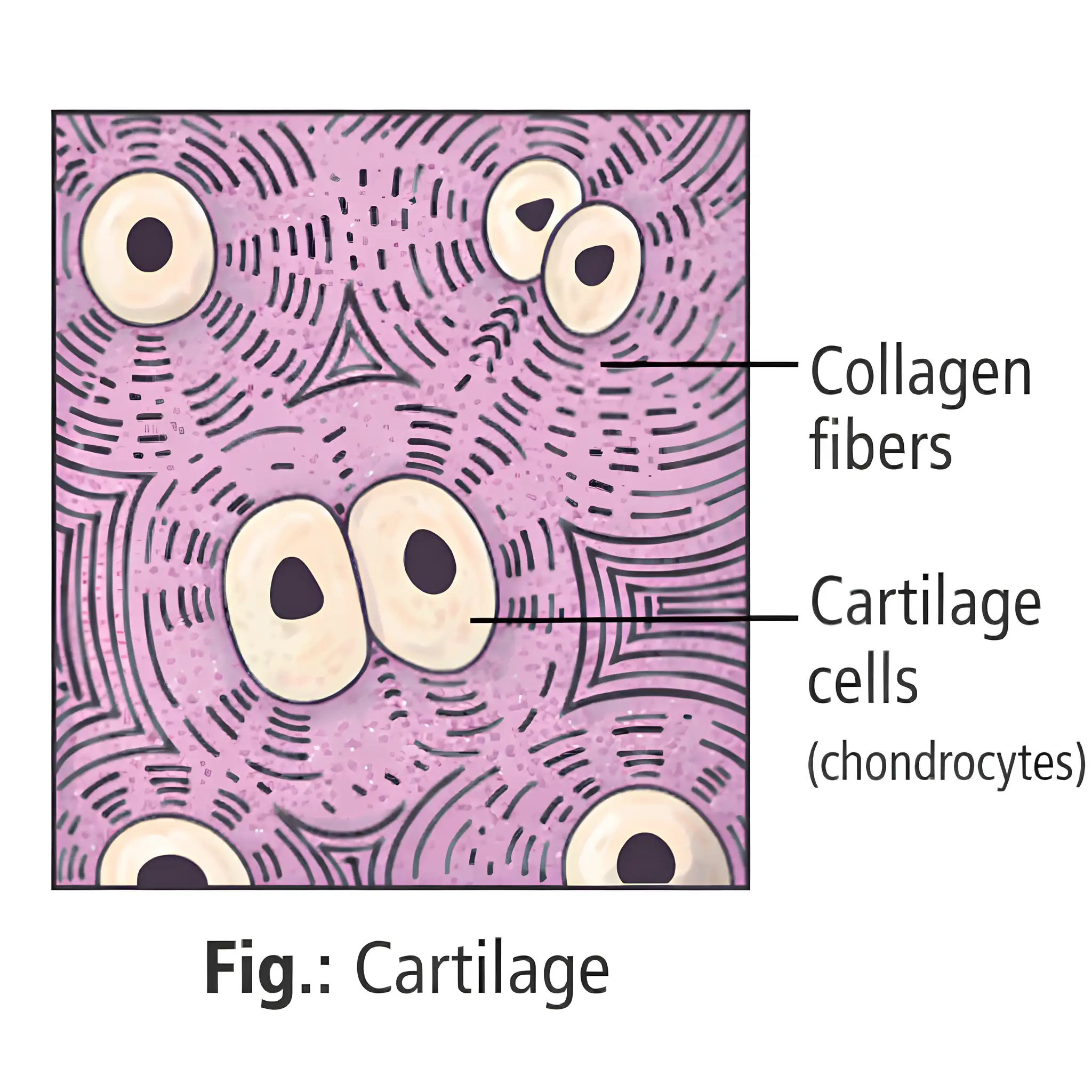
- Soft skeletal tissue with chondrocytes in lacunae.
- Matrix is solid, pliable, resists compression.
- Most embryonic cartilage replaced by bone in adults.
- Types: Hyaline (common, joints, trachea), White fibrous (strongest, intervertebral discs), Yellow elastic (ear, epiglottis).
Bone
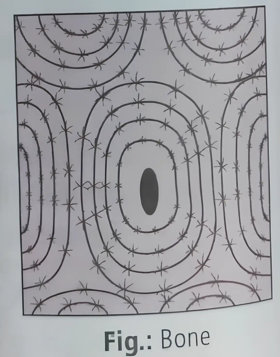
- Osteocytes in lacunae; matrix rich in calcium salts and collagen.
- Provides body frame, support, protection.
- Limb bones aid movement; red marrow produces blood cells.
- Osteon (Haversian system) = lamellae + lacunae + Haversian canal; absent in spongy bone.
Blood
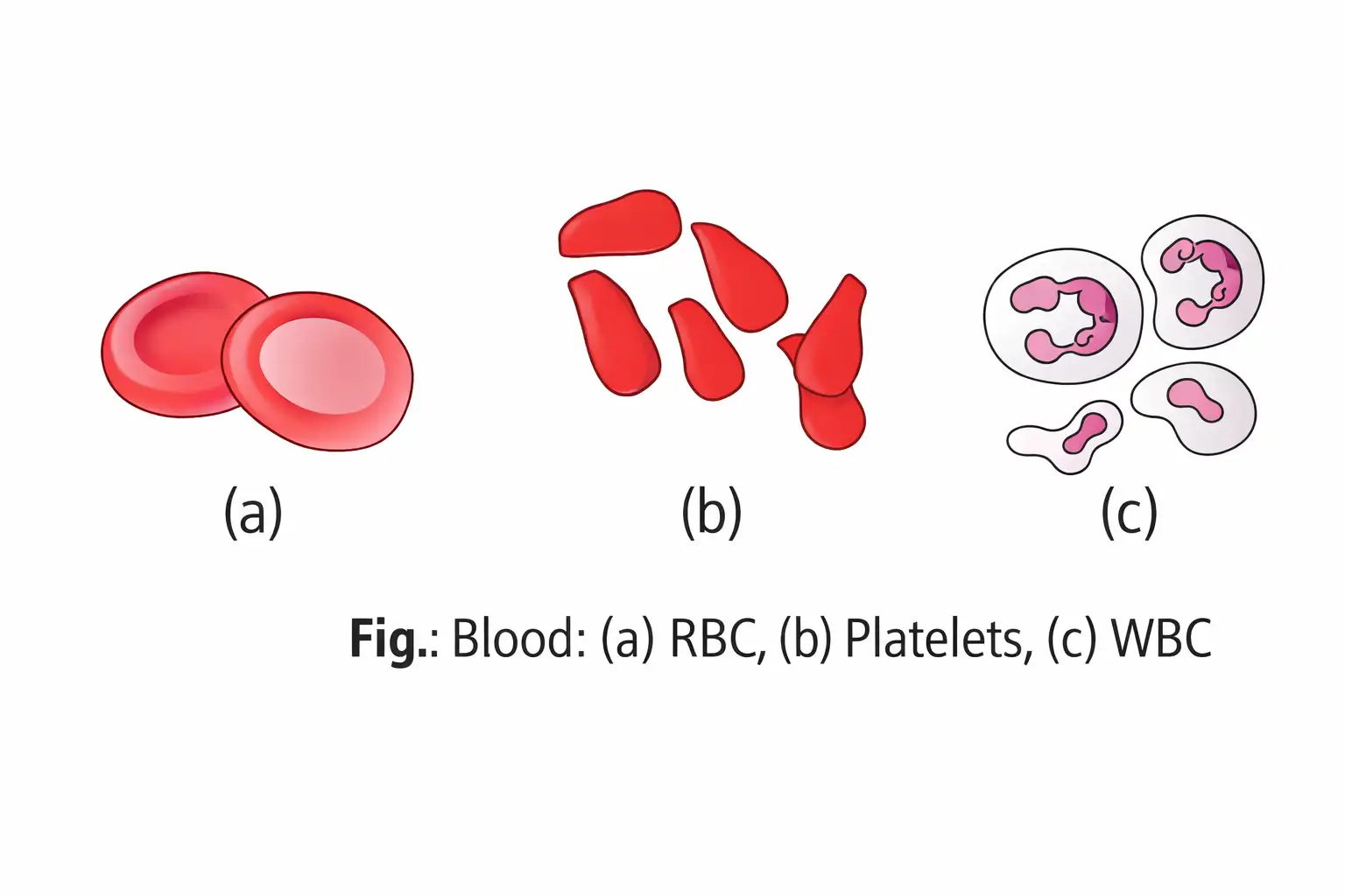
- Fluid connective tissue with plasma, RBCs, WBCs, platelets.
- Transports substances throughout the body.
- RBCs carry oxygen; platelets aid clotting; WBCs fight infection.
Memory Aids
- Cartilage = flexible support; Bone = hard support; Blood = transport.
- Osteon is the functional unit of compact bone.
Quick Checkpoints
- What are the types of cartilage and their locations?
- Describe the structure of an osteon.
- What are the components of blood and their functions?
Muscular Tissue
Structure and Function
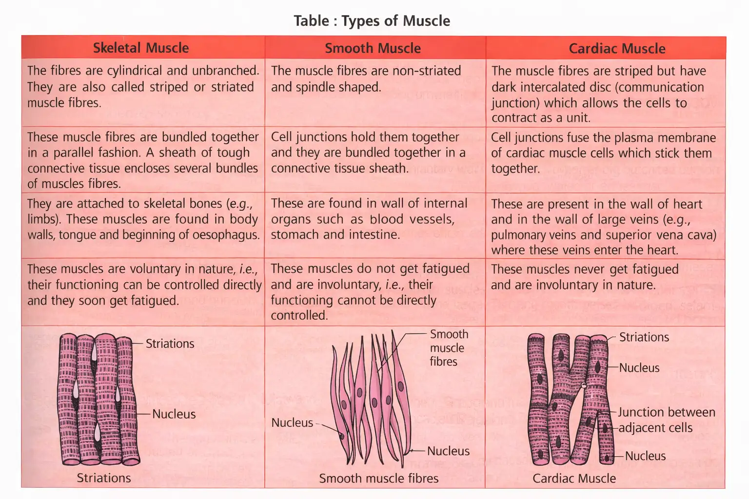
- Made of long cylindrical fibers with myofibrils in sarcoplasm.
- Fibers contract and relax to produce movement or maintain posture.
- Three types: Skeletal (voluntary, striated), Smooth (involuntary, non-striated), Cardiac (involuntary, striated with intercalated discs).
- Skeletal muscles attach to bones; smooth muscles in internal organs; cardiac muscle in heart.
Memory Aids
- Skeletal = voluntary, striated; Smooth = involuntary, no striations; Cardiac = involuntary, striated with discs.
Quick Checkpoints
- What are the three types of muscle tissue?
- Where is smooth muscle found?
- What special features does cardiac muscle have?
Neural Tissue
Structure and Function
- Controls body's response to stimuli.
- Neurons are the main units; consist of cyton (cell body), dendrites, and axon.
- Axon ends in terminal arborisations; axon/dendrite with sheath called nerve fiber.
- Neuroglial cells support and protect neurons; >50% of neural tissue volume.
- Nerve fibers: medullated (with myelin sheath, nodes of Ranvier) and non-medullated (without sheath).
- Electrical impulses travel along neuron membrane when stimulated.
Memory Aids
- Neuron = cyton + dendrites + axon; neuroglia support neurons.
- Myelin sheath speeds impulse; nodes of Ranvier are gaps.
Quick Checkpoints
- What are the parts of a neuron?
- Difference between medullated and non-medullated nerve fibers?
- Role of neuroglial cells?
Organ and Organ System
Overview
- Basic tissues form organs (e.g., heart has epithelial, connective, muscular, neural tissues).
- Organs combine to form organ systems; organ systems form an organism.
- Organisation ensures efficient, coordinated cell activities.
- Morphology: Study of external features of animals.
- Anatomy: Study of internal organ morphology.
Memory Aids
- Organ = Tissues combined; Organ system = Organs combined.
- Morphology = External form; Anatomy = Internal structure.
Quick Checkpoints
- What tissues form an organ?
- Difference between morphology and anatomy?
- Why is organ organisation important?
Cockroach
General Characteristics
- Scientific name: Periplaneta americana.
- Class: Insecta; Phylum: Arthropoda.
- Brown or black, nocturnal, omnivorous.
- Body divided into head, thorax, abdomen; covered by chitinous exoskeleton.
- Exoskeleton plates called sclerites (tergites dorsal, sternites ventral) joined by flexible membranes.
- Head triangular, fused from 6 segments, flexible neck, with compound eyes and antennae.
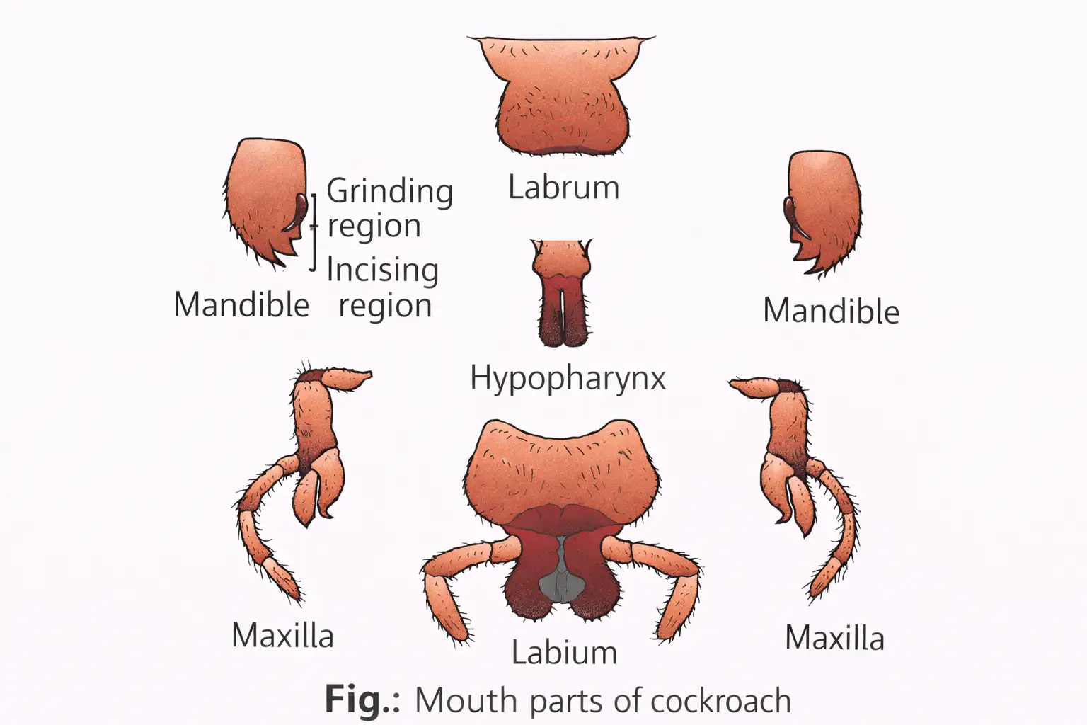
- Mouthparts chewing type: labrum (upper lip), labium (lower lip), maxillae, mandibles, hypopharynx (tongue-like).
- Thorax has prothorax, mesothorax, metathorax; each with a pair of legs.
- Two pairs of wings: forewings (opaque, leathery), hindwings (transparent, membranous for flight).
Mouthparts Function
- Mandibles: strong jaws for cutting and crushing food; grinding and incising regions.
- Maxillae: hold and manipulate food.
- Labrum: upper lip, holds food in place.
- Hypopharynx: inside mouth, mixes food with saliva.
- Labium: lower lip, holds food during eating.
Memory Aids
- Mandibles = jaws; Maxillae = food holders; Labrum = upper lip; Labium = lower lip; Hypopharynx = tongue.
Quick Checkpoints
- What are the main mouthparts of cockroach?
- Function of mandibles and maxillae?
- Role of hypopharynx?
Morphology
Body Structure
- Body segmented into head, thorax, abdomen.
- Covered by hard chitinous exoskeleton with sclerites (tergites dorsal, sternites ventral) joined by flexible membranes.
- Head triangular, fused from 6 segments, flexible neck, with compound eyes and antennae.
- Mouthparts: labrum (upper lip), labium (lower lip), maxillae, mandibles, hypopharynx (tongue-like).
- Thorax: prothorax, mesothorax, metathorax; each segment has a pair of walking legs with segmented podomeres.
- Wings: two pairs from meso- and metathorax; forewings opaque and leathery, hindwings transparent and membranous for flight.
Memory Aids
- Head = 6 fused segments; Thorax = 3 parts with legs; Abdomen = 10 segments.
- Forewings cover hindwings at rest.
Quick Checkpoints
- How is cockroach body segmented?
- What are sclerites?
- Describe the wings and their function.
Anatomy
Body Segments and Sexual Differences
- Abdomen has 10 segments in both sexes.
- Female 7th sternum boat-shaped; 7th-9th sterna form brood pouch with female gonopore, spermathecal pores, collaterial glands.
- Male genital pouch at hind abdomen, bounded by 9th-10th terga and 9th sternum; contains anus, male genital pore, gonapophysis.
- Males have short anal styles; both sexes have anal cerci on 10th segment.
Memory Aids
- Female brood pouch = 7th-9th sterna; Male genital pouch = 9th-10th terga + 9th sternum.
- Anal cerci present in both sexes; anal styles only in males.
Quick Checkpoints
- How many abdominal segments in cockroach?
- Differences in male and female genital structures?
- What are anal cerci and anal styles?
Digestive system
Structure and Function
- Alimentary canal divided into foregut, midgut, hindgut.
- Foregut: Mouth → pharynx → oesophagus → crop (stores food) → gizzard (proventriculus) with 6 chitinous teeth for grinding.
- Midgut: Short, narrow, coiled; junction with foregut has 6-8 hepatic caecae secreting digestive juice.
- Midgut-hindgut junction has 100-150 Malpighian tubules removing excretory products.
- Hindgut: Ileum, colon, rectum with 6 rectal glands; opens via anus; more permeable to water than foregut.
- Foregut and hindgut ectodermal, lined by cuticle; midgut endodermal, no cuticle.
Memory Aids
- Crop stores food; Gizzard grinds food.
- Hepatic caecae = digestive juice; Malpighian tubules = excretion.
- Foregut/hindgut lined with cuticle; midgut not.
Quick Checkpoints
- What are the three regions of alimentary canal?
- Function of crop and gizzard?
- Role of hepatic caecae and Malpighian tubules?
Blood vascular system
Features
- Open type system; vessels open into haemocoel.
- Haemolymph bathes visceral organs; consists of colourless plasma and haemocytes.
- No respiratory pigment; does not aid respiration.
- Heart is elongated muscular tube with funnel-shaped chambers and ostia; pumps blood anteriorly.
- Heart is neurogenic.
Memory Aids
- Open system with haemocoel.
- Haemolymph lacks respiratory pigment.
- Heart has ostia and is neurogenic.
Quick Checkpoints
- Type of blood vascular system?
- Composition and function of haemolymph?
- What is neurogenic heart?
Respiratory system
Structure and Function
- Network of tracheae opening through 10 pairs of spiracles on body sides.
- Spiracles guarded by bristles to prevent dirt entry.
- Tracheal tubes carry oxygen to tissues; subdivide into tracheoles for gas exchange by diffusion.
- Most CO2 exits through body cuticle, little through spiracles.
Memory Aids
- 10 pairs of spiracles with bristles.
- Tracheoles = site of gas exchange.
- CO2 mostly leaves via cuticle.
Quick Checkpoints
- How does cockroach breathe?
- Function of spiracles and tracheae?
- Pathway of CO2 exit?
Excretory system
Components and Function
- Excretion by Malpighian tubules lined with glandular and ciliated cells.
- Malpighian tubules absorb nitrogenous wastes, convert to uric acid; cockroach is uricotelic.
- Also reabsorb salts and water.
- Fat body, nephrocytes, uricose glands assist excretion.
Memory Aids
- Malpighian tubules = uric acid excretion.
- Insects are uricotelic.
- Fat body and nephrocytes aid excretion.
Quick Checkpoints
- What excretes nitrogenous wastes?
- Why are insects uricotelic?
- Other excretory aids?
Nervous system
Structure and Features
- Comprises central, peripheral, sympathetic systems.
- Fused, segmental ganglia joined by paired longitudinal connectives ventrally.
- 9 ganglia: 3 thoracic, 6 abdominal.
- Brain = bilobed supra-oesophageal ganglion; supplies antennae and compound eyes.
- Compound eyes have ~2000 hexagonal ommatidia; provide mosaic nocturnal vision (high sensitivity, low resolution).
- Other sense organs: antennae, maxillary palps, labial palps, anal cerci.
Memory Aids
- 9 ganglia: 3 thorax, 6 abdomen.
- Brain = supra-oesophageal ganglion.
- Compound eyes = mosaic vision with ommatidia.
Quick Checkpoints
- Number and location of ganglia?
- Function of supra-oesophageal ganglion?
- What is mosaic vision?
Reproductive system
Male and Female Structures
- Dioecious: sexes separate.
- Male: testes in 4th-6th abdominal segments, 3-lobed with follicles.
- Vas deferens opens into ejaculatory duct via seminal vesicles; stores sperms as spermatophores.
- Accessory mushroom-shaped gland in 6th-7th segments.
- External genitalia: male gonapophyses (phallomeres).
- Female: ovaries in 2nd-6th segments with ovarioles containing developing ova.
- Oviducts unite into median oviduct opening into genital chamber.
- Spermathecae in 6th segment open into genital chamber.
- Collaterial glands behind ovaries secrete ootheca (egg-case).
- Sperms transferred as spermatophores; fertilized eggs encased in reddish-brown oothecae.
- Females produce 9-10 oothecae, each with 14-16 eggs.
- Development is paurometabolous via nymphal stage; nymph moults ~13 times to adult.
- Some species are pests, contaminating food and spreading bacterial diseases.
Memory Aids
- Dioecious = separate sexes.
- Male: testes → vas deferens → ejaculatory duct → gonopore.
- Female: ovaries → oviduct → genital chamber; ootheca from collaterial glands.
- Development: paurometabolous with moulting.
Quick Checkpoints
- What does dioecious mean?
- Describe male reproductive tract.
- What forms the ootheca?
- Type of development in cockroach?
Frog
Characteristics
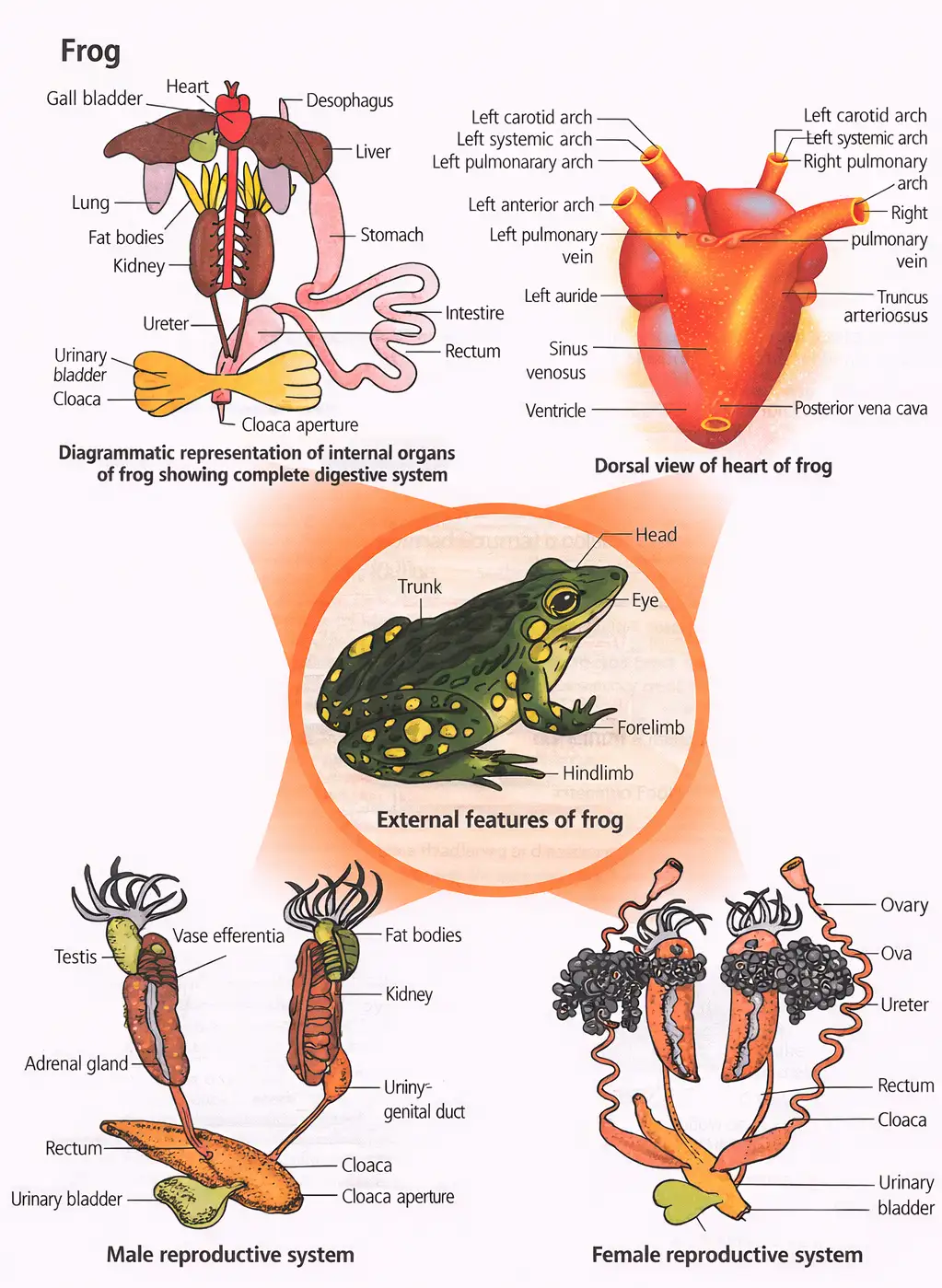
- Class Amphibia, Phylum Chordata; live in water and on land (amphibians).
- Common Indian frog: Rana tigrina.
- Cold-blooded (poikilotherms): body temperature varies with environment.
- Can change body color for camouflage (mimicry).
- Undergo aestivation (summer sleep) and hibernation (winter sleep) in extreme conditions.
Memory Aids
- Amphibians = live in both water and land.
- Poikilotherms = body temperature varies.
- Mimicry = protective coloration.
- Aestivation = summer sleep; Hibernation = winter sleep.
Quick Checkpoints
- What class and phylum does frog belong to?
- Define poikilotherms.
- What is mimicry in frogs?
- What are aestivation and hibernation?
Morphology
External Features
- Dorsal color: olive green with dark spots; ventral side pale yellow.
- Skin moist, smooth, slippery; absorbs water, does not drink.
- Body parts: head and trunk; no neck or tail.
- Nostrils above mouth; bulged eyes with nictitating membrane.
- Tympanum (ear) on sides of eyes for sound reception.
- Forelimbs: 4 digits; hindlimbs: 5 digits, muscular, webbed for swimming.
- Sexual dimorphism: males have vocal sacs and copulatory pads; males darker and slender.
Memory Aids
- Skin absorbs water, not drinking.
- Hindlimbs larger, webbed for swimming.
- Male vocal sacs and copulatory pads for mating.
Quick Checkpoints
- What are the body divisions of a frog?
- How do frogs absorb water?
- Describe sexual dimorphism in frogs.
- Function of tympanum?
Anatomy
Digestive System
- Short alimentary canal; frogs are carnivores.
- Mouth → buccal cavity → pharynx → oesophagus → stomach (cardiac and pyloric parts).
- Small intestine: duodenum (digestion) and ileum (absorption).
- Rectum opens into cloaca.
- Digestive glands: liver (bile), pancreas (pancreatic juice), gastric and intestinal glands.
- Food captured by bilobed tongue; digestion by HCl and gastric juices.
- Chyme passes to duodenum; bile emulsifies fats; enzymes digest carbs and proteins.
- Absorption by villi and microvilli in intestine.
- Undigested waste expelled via cloaca.
Respiratory System
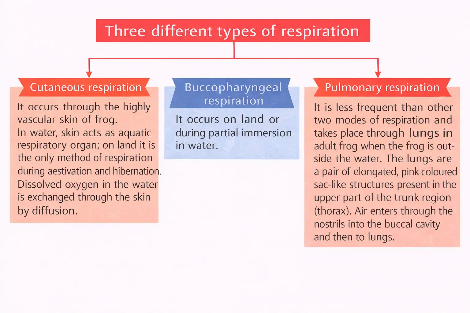
- Three types of respiration: cutaneous (skin), buccopharyngeal (mouth and throat), pulmonary (lungs).
- Cutaneous respiration mainly in water; pulmonary on land via nostrils to lungs.
Circulatory System
- Closed circulatory system: heart, blood vessels, blood; lymphatic system present.
- Heart: 3 chambers (2 atria, 1 ventricle), covered by pericardium.
- Sinus venosus joins right atrium; ventricle opens into conus arteriosus.
- Arteries carry blood from heart; veins return blood to heart.
- Hepatic portal system (liver-intestine) and renal portal system (kidney-lower body) present.
- Blood: plasma + cells (nucleated RBCs with hemoglobin, WBCs, platelets).
- Blood transports nutrients, gases, water; heart pumps blood.
Excretory System
- Components: paired kidneys, ureters, cloaca, urinary bladder.
- Kidneys: dark red, bean-shaped, contain nephrons (uriniferous tubules).
- Male ureters act as urinogenital ducts; female ureters and oviduct open separately into cloaca.
- Urinary bladder thin-walled, opens into cloaca.
- Frog is ureotelic; excretes urea.
- Excretory wastes filtered from blood by kidneys.
Memory Aids
- Short intestine due to carnivorous diet.
- Three respiration types: skin, mouth/throat, lungs.
- Heart has 3 chambers; closed circulation.
- Ureotelic animal: excretes urea.
Quick Checkpoints
- What are the parts of frog's digestive system?
- Explain the three types of respiration in frogs.
- Describe the structure of frog's heart.
- What is the excretory system composed of?
Control and Co-ordination
Neural and Endocrine Systems
- Control system includes nervous and endocrine systems.
- Endocrine glands: pituitary, thyroid, parathyroid, thymus, pineal, pancreatic islets, adrenals, gonads.
- Nervous system: central (brain, spinal cord), peripheral (cranial, spinal nerves), autonomic (sympathetic, parasympathetic).
- Brain enclosed in cranium; divided into forebrain, midbrain, hindbrain.
- Ten pairs of cranial nerves arise from brain.
- Sense organs: touch, taste, smell, vision (eyes), hearing (tympanum and internal ears).
Memory Aids
- Brain parts: forebrain, midbrain, hindbrain.
- 10 pairs cranial nerves.
- Endocrine glands regulate body functions via hormones.
Quick Checkpoints
- What are the components of frog's nervous system?
- Name major endocrine glands in frogs.
- How many cranial nerve pairs are present?
- List the sense organs of frog.
Reproductive System
Male and Female Systems
- Male: paired yellowish testes, vasa efferentia, urinogenital ducts.
- Testes attached to kidneys by mesorchium.
- Vasa efferentia open into Bidder's canal, which connects to urinogenital duct opening into cloaca.
- Cloaca passes faeces, urine, and sperms.
- Female: paired ovaries, oviducts, cloaca.
- Mature female lays 2500-3000 ova at a time.
- Fertilisation is external in water.
- Development includes larval tadpole stage; metamorphosis forms adult.
Memory Aids
- Mesorchium connects testes to kidneys.
- External fertilisation in water.
- Tadpole undergoes metamorphosis.
Quick Checkpoints
- Describe male reproductive organs in frog.
- How does fertilisation occur?
- What is metamorphosis?
- Number of ova laid by female frog?
Economic Importance
Benefits to Humans and Ecosystem
- Frogs eat insects, protecting crops.
- Maintain ecological balance as part of food chain and web.
- Frog legs consumed as food in some countries.
Memory Aids
- Frogs control insect population.
- Important link in ecosystem food chains.
- Edible frog legs in some cultures.
Quick Checkpoints
- How do frogs benefit agriculture?
- What role do frogs play in ecosystems?
- Are frogs used as food?
NEET Zoology — ALL CHAPTERS
1
Structural Organisation in Animals
2
Breathing and Exchange of Gases
3
Body Fluids and Circulation
4
Excretory Products and their Elimination
5
Locomotion and Movement
6
Neural Control and Coordination
7
Human Reproduction
8
Reproductive Health
9
Evolution
10
Microbes in Human Welfare
11
Organism and Population
12
Ecosystem
13
Biodiversity and Conservation