NEET Ug . NEET Zoology . Zoology . Body Fluids and Circulation
Chapter 3: Body Fluids and Circulation
Ch 3
NEET Zoology
NEET Ug
Introduction
Overview
- Living cells require nutrients, oxygen, and waste removal for healthy function.
- Complex organisms use body fluids for transport of substances.
- Blood is the primary transport fluid in higher organisms including humans.
- Lymph also aids in transport of certain substances.
Memory Aids
- Blood = main transport fluid in humans.
- Lymph = secondary transport fluid.
Quick Checkpoints
- What are the main transport fluids in humans?
- Why is a transport system essential?
Blood
Composition and Properties
- Blood is a mobile connective tissue with plasma (fluid matrix) and formed elements.
- Adult human blood volume: 5-5.5 litres, 30-35% of extracellular fluid.
- Blood is slightly alkaline with pH 7.4.
Memory Aids
- Blood volume ~5 litres in adults.
- pH ~7.4 (alkaline).
Quick Checkpoints
- What is the pH of blood?
- What percentage of extracellular fluid does blood constitute?
Plasma
Composition and Functions
- Plasma is a pale yellow, viscous, non-living intercellular fluid making up 55% of blood.
- 90-92% water; 6-8% plasma proteins including albumin, globulin, immunoglobulin, prothrombin, fibrinogen.
- Fibrinogen aids blood clotting; globulins aid defense; albumins maintain osmotic balance.
- Contains minerals (Na+, Ca2+, Mg2+, HCO3-, Cl-), glucose, amino acids, lipids in transit.
- Clotting factors are inactive in plasma; plasma without clotting factors is called serum.
Memory Aids
- Plasma = 55% blood, mostly water.
- Serum = plasma minus clotting factors.
Quick Checkpoints
- What is plasma made of?
- What is serum?
Formed Elements (Blood Corpuscles)
Types and Functions
- Formed elements: erythrocytes (RBCs), leucocytes (WBCs), platelets.
- They constitute about 45% of blood volume.
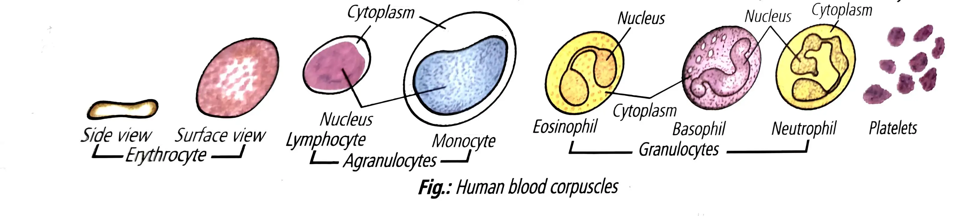
- Erythrocytes carry oxygen; biconcave, enucleated, formed in red bone marrow.
- Haemoglobin gives red color; lifespan ~120 days; formation called erythropoiesis.
- Iron, proteins, vitamin B12, folic acid essential for RBC formation.
- Spleen destroys RBCs; called graveyard of RBCs.
- Polycythemia = high RBC count; erythrocytopenia = low RBC count causing oxygen shortage; anemia = low haemoglobin.
- People in hills have more RBCs to adapt to low oxygen.
- Leucocytes (WBCs) are nucleated, colorless, 6000-8000 per cubic mm, short-lived.
- Two types: granulocytes (neutrophils, eosinophils, basophils) and agranulocytes (lymphocytes, monocytes).
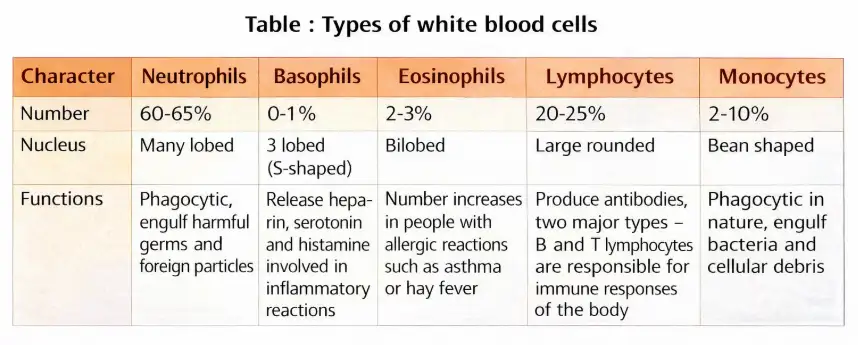
- Neutrophils most numerous; monocytes largest.
- Leucocyte formation called leucopoiesis; decrease called leukopenia; increase in leukemia.
- Platelets (thrombocytes) are cell fragments from megakaryocytes; 1.5-3.5 lakh per cubic mm.
- Platelets aid blood clotting; lifespan ~1 week; low count causes clotting disorders.
Memory Aids
- RBCs = oxygen carriers, biconcave, no nucleus.
- WBCs = immune defense, nucleated.
- Platelets = clotting, cell fragments.
Quick Checkpoints
- What are the three formed elements of blood?
- Where are RBCs formed?
- What causes anemia?
- Types of leucocytes?
- Role of platelets?
Blood Groups
ABO Blood Grouping
- Discovered by Karl Landsteiner (1900); AB group by Dacastello and Sturli (1902).
- Controlled by gene I with alleles Iᴬ, Iᴮ, Iᴼ.
- Iᴬ and Iᴮ produce A and B antigens on RBC surface; antibodies present in plasma.
- Donor and recipient blood groups must match to avoid RBC clumping.
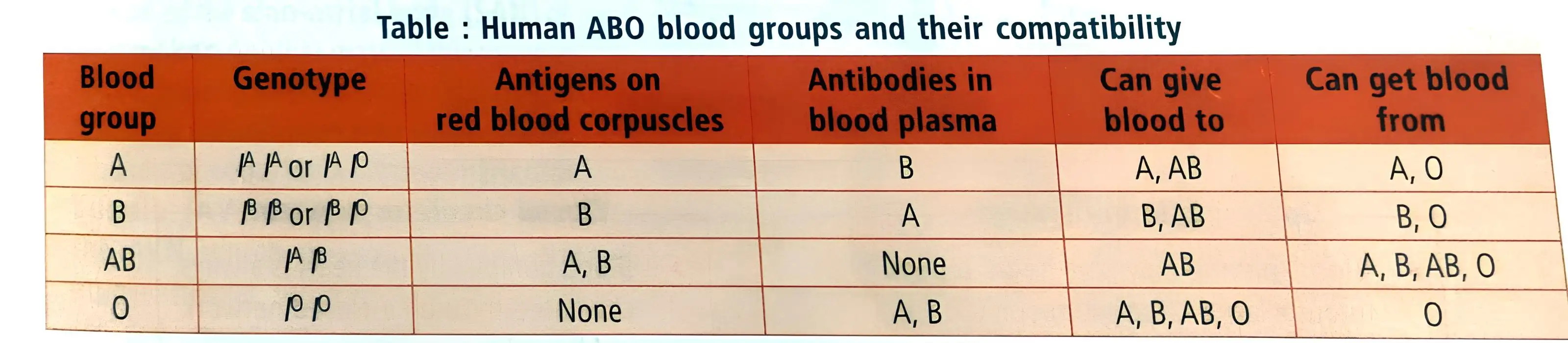
- Group O = universal donor; Group AB = universal recipient.
Rh Grouping
- Rh antigen discovered by Landsteiner and Wiener (1940) in Rhesus monkey.
- Present in ~80% humans; Rh+ve have antigen, Rh-ve lack it.
- Rh-ve exposed to Rh+ve blood form antibodies; matching needed before transfusion.
- Rh incompatibility in pregnancy causes erythroblastosis fetalis (fetal RBC destruction, anemia, jaundice).
- Prevented by anti-Rh antibodies given to mother after first delivery.
Memory Aids
- ABO: A and B antigens; O no antigen.
- Rh+: antigen present; Rh-: antigen absent.
- O = universal donor; AB = universal recipient.
Quick Checkpoints
- What alleles control ABO blood groups?
- Who is universal donor and recipient?
- What causes erythroblastosis fetalis?
Coagulation of Blood
Process and Importance
- Blood clotting stops bleeding after injury to blood vessels.
- Clot is a fibrin network trapping blood cells, forming a seal.
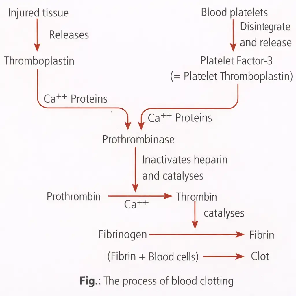
- Steps: injury releases thromboplastin; platelets release platelet factor 3.
- Thromboplastin + platelet factor 3 + Ca2+ form prothrombinase.
- Prothrombinase converts prothrombin to thrombin.
- Thrombin converts fibrinogen to fibrin, forming clot.
- Clotting time: 3-8 minutes.
- Vitamin K needed for prothrombin synthesis.
- Anticoagulants (heparin, oxalates) prevent clotting.
Memory Aids
- Clotting = thromboplastin → prothrombinase → thrombin → fibrin.
- Vitamin K essential for clotting.
- Heparin = anticoagulant.
Quick Checkpoints
- What triggers blood clotting?
- Role of thrombin?
- Why is vitamin K important?
Lymph (Tissue Fluid)
Composition and Characteristics
- Mobile connective tissue with lymph plasma and lymph corpuscles.
- Lymph plasma: Similar to blood plasma but with fewer proteins, less calcium and phosphorus, and higher glucose.
- Collected and drained by the lymphatic system into major veins.
Cellular Components and Functions
- Lymph corpuscles: Amoeboid leucocytes, mainly lymphocytes; no erythrocytes or platelets.
- Transports nutrients, hormones, and fats (absorbed via lacteals in intestinal villi).
- Acts as a transporter between body cells and blood, carrying wastes like carbon dioxide to blood.
- Destroys microorganisms and foreign particles in lymph nodes.
Memory Aids
- Lymph = Plasma + Corpuscles (mostly lymphocytes).
- Fats absorbed via lacteals → transported by lymph.
- Lymphatic system drains fluid back to veins.
Quick Checkpoints
- What are the main components of lymph?
- How does lymph plasma differ from blood plasma?
- Which cells are present and absent in lymph?
- What is the role of lymph in fat absorption?
- How does lymph contribute to immune defense?
Circular Pathways
Types of Circulatory Systems
- Open system: Blood leaves vessels, fills sinuses; found in arthropods, mollusks.
- Closed system: Blood stays in vessels, circulates continuously; found in annelids, chordates.
- Closed system allows precise regulation of fluid flow.
- Evolution of vertebrate hearts:
- 2-chambered: 1 atrium, 1 ventricle; single circulation in fishes; pumps deoxygenated blood to gills.
- 3-chambered: 2 atria, 1 ventricle; incomplete double circulation in amphibians, reptiles; mixed blood pumped.
- 4-chambered: 2 atria, 2 ventricles; double circulation in crocodiles, birds, mammals; no blood mixing.
Memory Aids
- Open = blood leaves vessels; Closed = blood stays in vessels.
- 2-chambered = fish, single loop; 3-chambered = amphibians/reptiles, mixed blood; 4-chambered = birds/mammals, separate loops.
Quick Checkpoints
- What distinguishes open from closed circulatory systems?
- How does blood flow differ in 2, 3, and 4-chambered hearts?
- Why is closed circulation more efficient?
Human Circulatory System
Components and Heart Structure
- Also called blood vascular system: muscular heart, closed vessels (arteries, veins), blood.
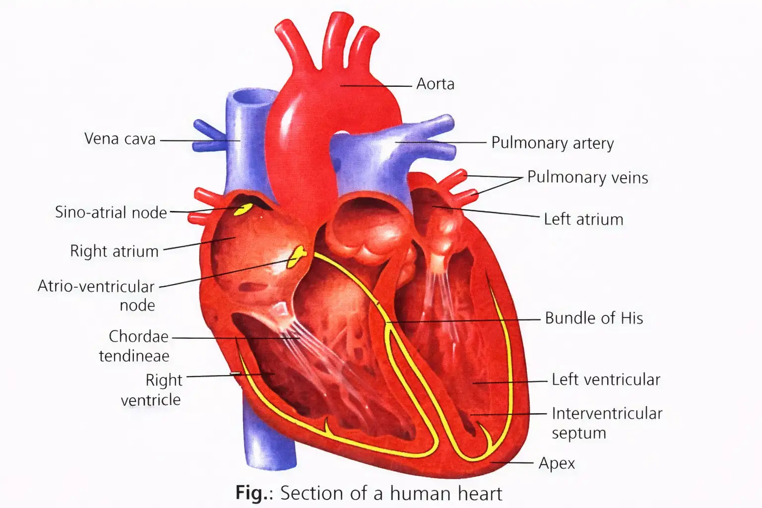
- Heart: four-chambered, mesodermal, in thoracic cavity, left tilted, fist-sized.
- Enclosed in pericardium with pericardial cavity and fluid.
- Right and left atria separated by interatrial septum; ventricles by interventricular septum.
- Right atrium receives deoxygenated blood via superior and inferior vena cava.
- Left atrium receives oxygenated blood from lungs via pulmonary veins.
- Left ventricle thicker to pump blood at higher pressure.
- Atrioventricular septum separates atrium and ventricle on same side with valves:
- Bicuspid (mitral) valve on left side.
- Tricuspid valve on right side.
- Chordae tendineae connect valve flaps to papillary muscles.
- Semilunar valves at pulmonary artery and aorta openings prevent backflow.
- Heart muscle includes nodal tissue: sino-atrial node (SAN) as pacemaker, atrioventricular node (AVN) as pacesetter.
- AV bundle (bundle of His) and Purkinje fibers conduct impulses for rhythmic contraction.
- Heartbeat types:
- Neurogenic: nerve impulse initiated (annelids, arthropods).
- Myogenic: muscle initiated (molluscs, vertebrates including humans).
Memory Aids
- SAN = pacemaker; AVN = pacesetter.
- Left ventricle thicker = higher pressure pump.
- Bicuspid = 2 cusps; Tricuspid = 3 cusps.
Quick Checkpoints
- What separates atria and ventricles?
- Which valves prevent backflow into atria?
- What is the role of nodal tissue?
- Difference between neurogenic and myogenic heartbeat?
Cardiac Cycle
Stages and Events
- Cardiac cycle = systole (contraction) + diastole (relaxation) of atria and ventricles.
- Stages:
- Joint diastole: all chambers relaxed, AV valves open, semilunar valves closed, ventricles fill.
- Atrial systole: atria contract, increase ventricular filling by 30%.
- Ventricular systole: ventricles contract, AV valves close (first heart sound 'lub'), semilunar valves closed initially.
- Ventricular diastole: semilunar valves open, blood ejected, ventricles relax, semilunar valves close (second heart sound 'dub').
- Complete ventricular diastole: ventricles relax, AV valves open, cycle repeats.
- Cardiac output = heart rate × stroke volume (~5.5 L/min in humans).
- Duration of cardiac cycle = 0.8 seconds.
- Heart sounds: 'lub' (0.15 s, 25-45 Hz), 'dub' (0.12 s, 50 Hz).
Memory Aids
- Lub = AV valves closing; Dub = semilunar valves closing.
- Atrial systole = 0.1 s; Ventricular systole = 0.3 s; Joint diastole = 0.4 s.
- Cardiac output depends on heart rate and stroke volume.
Quick Checkpoints
- What happens during atrial systole?
- Which valves produce the first and second heart sounds?
- How is cardiac output calculated?
- Duration of one cardiac cycle?
Electrocardiogram (ECG)
Basics and Components
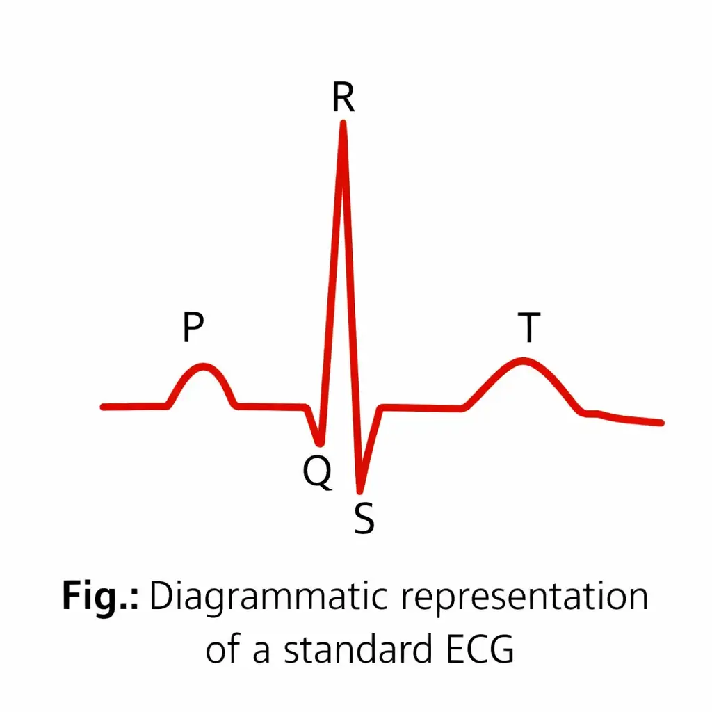
- ECG records electrical activity of cardiac muscles using electrocardiograph.
- Standard ECG uses three leads: two wrists, one left ankle.
- Normal ECG waves:
- P wave: atrial depolarisation (contraction), caused by SA node.
- QRS complex: ventricular depolarisation (contraction), starts with Q (down), R (up), S (down).
- T wave: ventricular repolarisation (relaxation), dome-shaped.
- End of T wave marks end of systole.
- Heart rate can be determined by counting QRS complexes per time.
- Abnormal ECG shapes indicate heart diseases.
Memory Aids
- P wave = atrial contraction.
- QRS complex = ventricular contraction.
- T wave = ventricular relaxation.
Quick Checkpoints
- What does the P wave represent?
- Which wave indicates ventricular contraction?
- How is heart rate measured from ECG?
- What does an abnormal ECG indicate?
Double Circulation
Blood Vessels
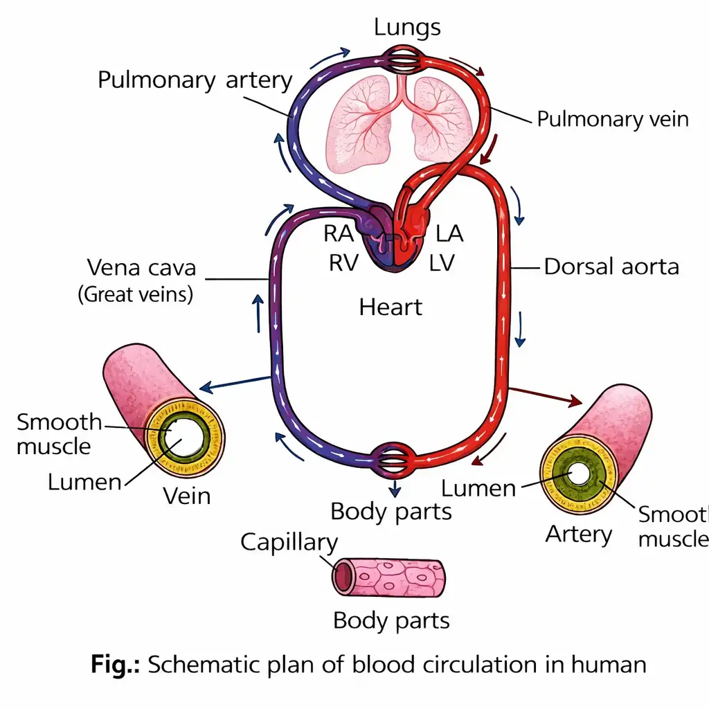
- Three types: arteries, veins, capillaries.
- Arteries: Elastic vessels carrying oxygenated blood from heart to tissues; pulmonary artery carries deoxygenated blood.
- Veins: Carry deoxygenated blood to heart; pulmonary vein carries oxygenated blood from lungs to left atrium; veins have valves to prevent backflow.
- Capillaries: Connect arteries and veins in tissues.
- Walls of arteries and veins have three layers:
- Tunica intima: Innermost, squamous endothelium + elastic membrane.
- Tunica media: Middle, smooth muscle + elastic fibers; thinner in veins.
- Tunica externa: Outer fibrous connective tissue with collagen.
Double Circulation Pathways
- Blood passes twice through heart in separate routes per cycle.
- Consists of pulmonary circulation and systemic circulation.
Pulmonary Circulation
- Deoxygenated blood pumped from right ventricle to lungs via pulmonary trunk and arteries.
- Blood gets oxygenated in lungs.
- Oxygenated blood returns to left atrium via pulmonary veins.
Systemic Circulation
- Oxygenated blood pumped from left ventricle to body through arteries, arterioles, capillaries.
- Deoxygenated blood collected by venules, veins, vena cava, returns to right atrium.
- Delivers O2, nutrients; removes CO2, wastes.
Special Circulations
- Hepatic portal system: Connects digestive tract to liver.
- Coronary circulation: Supplies blood to cardiac muscle.
Memory Aids
- Arteries away, veins to heart (except pulmonary vessels).
- Pulmonary artery = deoxygenated; pulmonary vein = oxygenated.
- Double circulation = two heart passes per blood cycle.
- Hepatic portal = digestive tract to liver.
- Coronary = heart muscle blood supply.
Quick Checkpoints
- What are the three types of blood vessels and their functions?
- Which artery carries deoxygenated blood?
- Describe the layers of artery and vein walls.
- Explain pulmonary circulation steps.
- Explain systemic circulation steps.
- What is the hepatic portal system?
- What is coronary circulation?
Regulation of Cardiac Activity
Autoregulation by Nodal Tissue
- Heart's normal activity controlled by specialized muscles called nodal tissue.
- Heart is myogenic: it generates its own impulses without external stimuli.
- Medulla oblongata contains a neural center that modulates heart function via the autonomic nervous system (ANS).
Neural and Hormonal Control
- Sympathetic nerves (ANS) increase heart rate, ventricular contraction strength, and cardiac output.
- Parasympathetic nerves (ANS) decrease heart rate, slow action potential conduction, and reduce cardiac output.
- Adrenal medullary hormones also enhance cardiac output.
Memory Aids
- Myogenic = Heart beats by itself.
- Sympathetic = Speed up heart.
- Parasympathetic = Slow down heart.
- Medulla oblongata = Cardiac control center.
Quick Checkpoints
- What does myogenic mean in cardiac activity?
- How does the sympathetic nervous system affect the heart?
- Role of parasympathetic nerves in heart regulation?
- Which hormones increase cardiac output?
Disorders of Circulatory System
Hypertension and Blood Pressure
- Hypertension is high blood pressure above 140/90 mmHg, harming heart, brain, kidneys, and eyes.
- Blood pressure is the force exerted by blood on artery walls.
- Systolic pressure (120 mmHg) is pressure when left ventricle contracts.
- Diastolic pressure (80 mmHg) is pressure when heart rests during diastole.
- Blood pressure is expressed as BP = 120/80 mmHg.
- Difference between systolic and diastolic pressure is pulse pressure.
- Measured using a sphygmomanometer.
Memory Aids
- Normal BP: 120/80 (Systolic/Diastolic)
- Hypertension if BP > 140/90
- Pulse pressure = Systolic - Diastolic
Quick Checkpoints
- What defines hypertension?
- Difference between systolic and diastolic pressure?
- Device used to measure blood pressure?
Atherosclerosis and Coronary Artery Disease
Causes and Effects
- Atherosclerosis/CAD is lipid, fat, calcium, and fibrous tissue buildup forming atheromatous plaques in arteries.
- Plaques can block arteries, reducing blood supply to heart muscle, causing heart attack or stroke.
- Arteriosclerosis is hardening and thickening of arteries due to calcium and cholesterol deposits.
- Hardened arteries lose elasticity and may rupture, leading to heart attack or death.
Memory Aids
- Atherosclerosis = Plaque buildup in arteries
- Arteriosclerosis = Artery hardening
- CAD affects coronary arteries causing heart issues
Quick Checkpoints
- What forms atheromatous plaques?
- Difference between atherosclerosis and arteriosclerosis?
- Consequences of artery blockage?
Angina
Definition and Symptoms
- Angina pectoris is acute chest pain due to insufficient oxygen to heart muscle.
- Affects all ages, more common in middle-aged and elderly.
- Caused by reduced blood flow to heart.
Memory Aids
- Angina = Chest pain from low oxygen
- Occurs due to blood flow issues
Quick Checkpoints
- What causes angina?
- Who is commonly affected?
Heart Failure
Definition and Symptoms
- Heart failure or congestive heart failure is when heart pumps blood ineffectively.
- Leads to lung congestion as a main symptom.
Memory Aids
- Heart failure = weak heart pump
- Causes lung congestion
Quick Checkpoints
- What is congestive heart failure?
- Main symptom of heart failure?
Cardiac Arrest
Definition
- Cardiac arrest is the complete stop of heart beating.
Memory Aids
- Cardiac arrest = Heart stops beating
Quick Checkpoints
- What happens in cardiac arrest?
Heart Attack
Definition
- Heart attack occurs when heart muscle is damaged due to inadequate blood supply.
Memory Aids
- Heart attack = Sudden heart muscle damage
Quick Checkpoints
- Cause of heart attack?
Human Heart Structure
Key Features
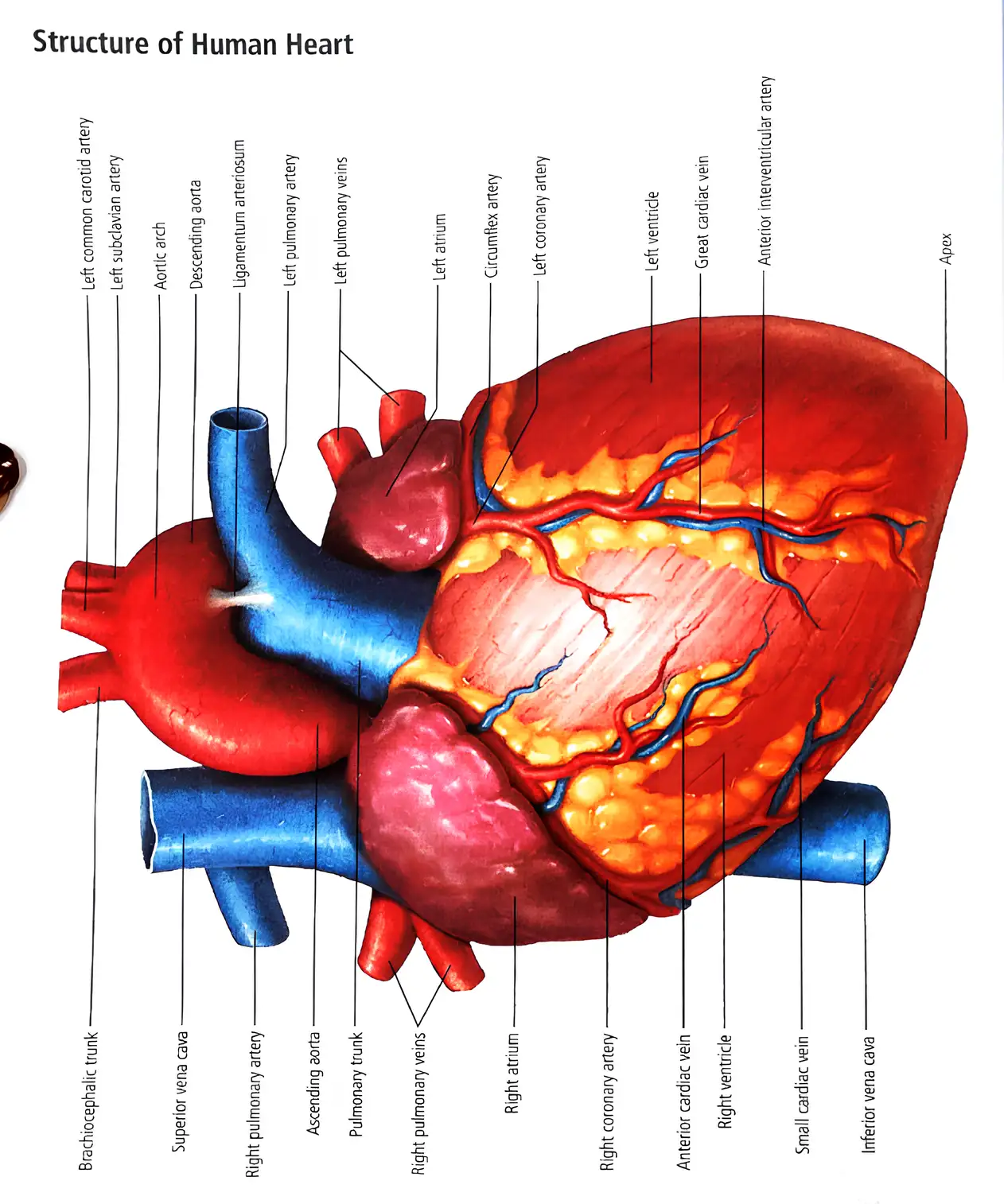
- Four chambers: left atrium, left ventricle, right atrium, right ventricle.
- Blood enters via superior and inferior vena cava.
- Right atrium receives oxygen-poor blood, sends to right ventricle.
- Right ventricle pumps blood to lungs via pulmonary artery.
- Oxygen-rich blood returns to left atrium via pulmonary veins.
- Left ventricle pumps blood to body through aorta.
- Heart supplied by coronary arteries and veins on surface.
- Smaller vessels circulate blood within heart muscle.
- Apex is the pointed end directed downwards.
Memory Aids
- Right side: deoxygenated blood; Left side: oxygenated blood
- Coronary arteries supply heart muscle
- Apex points downwards
Quick Checkpoints
- Name the four chambers of the heart.
- Path of oxygen-poor blood through the heart?
- Function of coronary arteries?
NEET Zoology — ALL CHAPTERS
1
Structural Organisation in Animals
2
Breathing and Exchange of Gases
3
Body Fluids and Circulation
4
Excretory Products and their Elimination
5
Locomotion and Movement
6
Neural Control and Coordination
7
Human Reproduction
8
Reproductive Health
9
Evolution
10
Microbes in Human Welfare
11
Organism and Population
12
Ecosystem
13
Biodiversity and Conservation