NEET Ug . NEET Zoology . Zoology . Human Reproduction
Chapter 7: Human Reproduction
Ch 7
NEET Zoology
NEET Ug
Introduction
Overview
- Humans reproduce sexually and are viviparous.
- Reproductive events: gametogenesis, insemination, fertilisation, zygote formation, blastocyst development, implantation, gestation, and parturition.
- Events occur after puberty with distinct male and female reproductive processes.
- Reproductive organs: primary (produce gametes and hormones) and secondary (conduct gametes, no gamete/hormone production).
- Secondary sexual characters are external features distinguishing sexes.
Memory Aids
- Primary = Produce gametes + hormones; Secondary = Conduct gametes only.
- Secondary sexual characters = External sex differences.
Quick Checkpoints
- What are the main reproductive events in humans?
- Define primary and secondary sex organs.
- What are secondary sexual characters?
Male Reproductive System
Structure and Location
- Located in the pelvis region.
- Includes paired testes, accessory ducts, accessory glands, and external genitalia.
- Testes are outside the abdominal cavity in the scrotum.
Memory Aids
- Testes in scrotum = outside abdomen for temperature control.
Quick Checkpoints
- Where is the male reproductive system located?
- What are the main components of the male reproductive system?
- Why are testes located outside the abdomen?
Scrotum
Function and Structure
- Connected to abdomen/pelvis by inguinal canals.
- Spermatic cord (artery, vein, nerve) passes through inguinal canal to testes.
- Maintains testes temperature at ~34°C, 2-2.5°C below body temperature.
- Temperature regulation via cremaster and dartos muscles adjusting scrotal position.
- Temperature receptors in scrotum control movement of testes towards/away from pelvis.
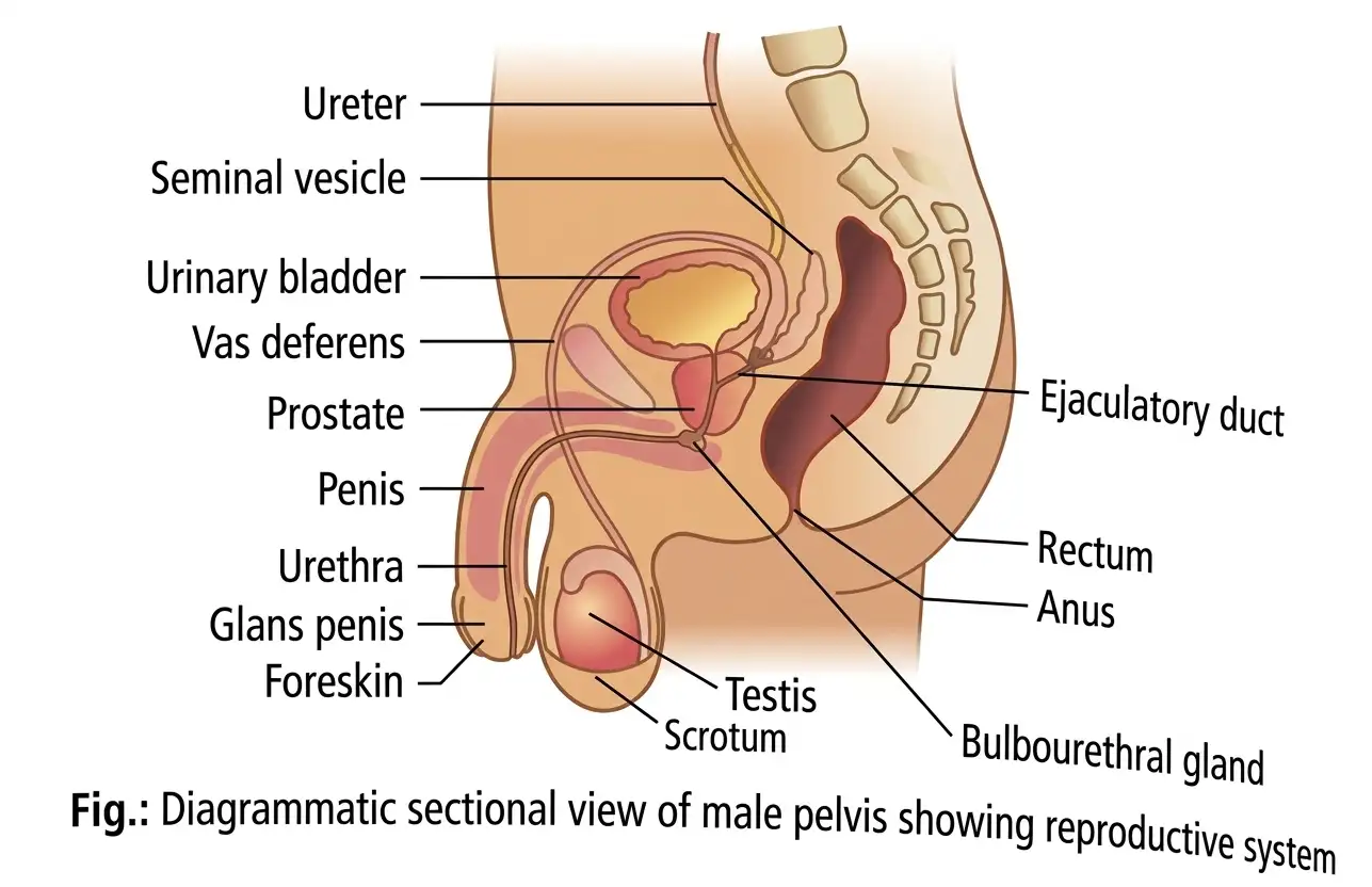
Memory Aids
- Scrotum = Temperature regulator for sperm production.
- Cremaster + Dartos muscles = Adjust testes position for heat control.
Quick Checkpoints
- How does the scrotum regulate testes temperature?
- What is the role of the spermatic cord?
- Why is testes temperature lower than body temperature?
Testis and Seminiferous Tubules
Structure and Function
- Testis: oval, 4-5 cm long, 2-3 cm wide; covered by tunica vaginalis, tunica albuginea, tunica vasculosa.
- Contains ~250 testicular lobules; each has 1-3 seminiferous tubules.
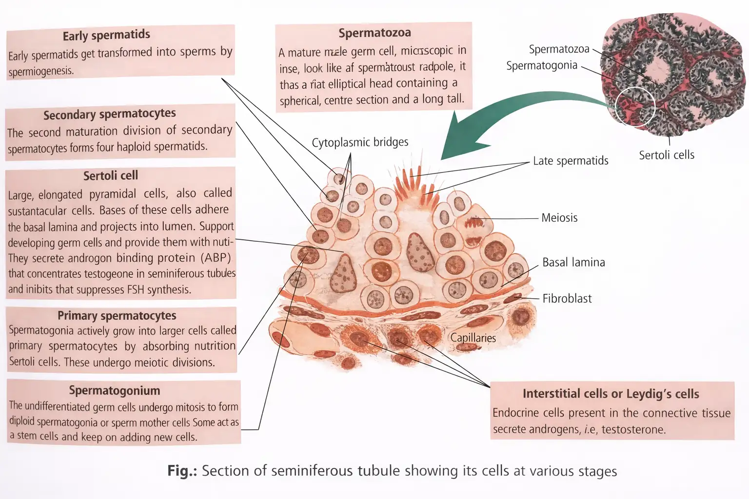
- Seminiferous tubules: site of spermatogenesis; lined by spermatogonia (male germ cells) and Sertoli cells (nutrition).
- Interstitial spaces contain blood vessels and Leydig (interstitial) cells producing androgens.
- Functions: sperm production and androgen secretion.
- Other immune cells present: lymphocytes, macrophages, mast cells.
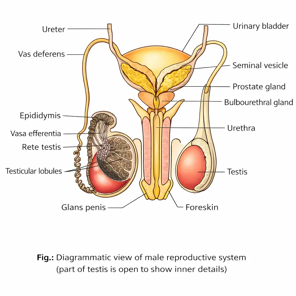
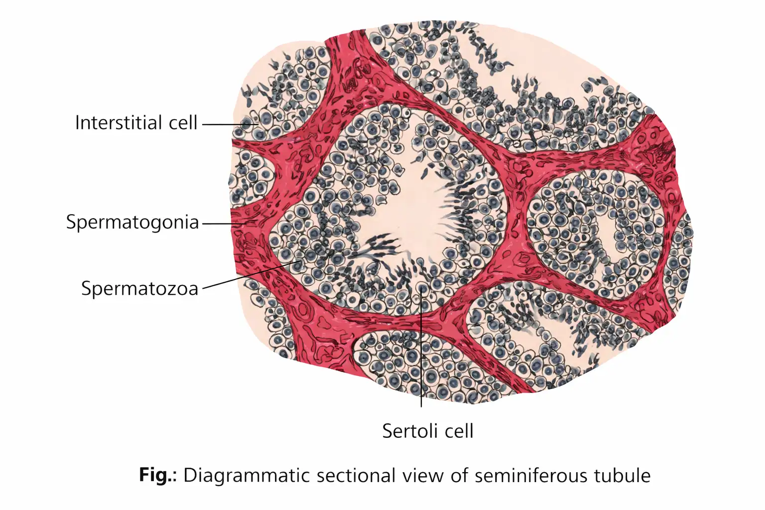
Memory Aids
- Seminiferous tubules = Sperm production site.
- Sertoli cells = Support and nourish sperm cells.
- Leydig cells = Produce testosterone.
Quick Checkpoints
- What are the layers covering the testis?
- Where does spermatogenesis occur?
- What cells produce testosterone?
Male Accessory Ducts
Components and Functions
- Include rete testis, vasa efferentia, epididymis, and vas deferens.
- Seminiferous tubules open into vasa efferentia via rete testis.
- Vasa efferentia lead to epididymis (posterior testis surface), where sperm mature and are stored.
- Epididymis divided into caput, corpus, and cauda regions.
- Vas deferens ascends to abdomen, loops over bladder, receives seminal vesicle duct, and opens into urethra as ejaculatory duct.
- Urethra extends from bladder through penis to urethral meatus (external opening).
Memory Aids
- Epididymis = Sperm maturation and storage.
- Vas deferens = Sperm transport to urethra.
Quick Checkpoints
- What is the path of sperm from seminiferous tubules to urethra?
- What are the parts of the epididymis?
- Where does the vas deferens open?
External Genitalia (Penis)
Structure and Function
- Penis: male copulatory organ and urinal duct.
- Contains three erectile tissues: two dorsal corpora cavernosa and one ventral corpus spongiosum.
- Corpus spongiosum surrounds penile urethra and forms glans penis at tip.
- Glans penis covered by prepuce (foreskin).
Memory Aids
- Corpora cavernosa = Erectile tissue for erection.
- Corpus spongiosum = Protects urethra during erection.
- Prepuce = Covers glans penis.
Quick Checkpoints
- What erectile tissues are present in the penis?
- What is the function of the corpus spongiosum?
- What is the prepuce?
Male Accessory Glands
Types and Secretions
- Include paired seminal vesicles, prostate gland, and paired bulbourethral (Cowper's) glands.
- Seminal vesicles: sac-like, near bladder base; ducts join vas deferens to form ejaculatory duct.
- Seminal vesicle secretion (60-70% semen volume): alkaline, contains fructose (energy), prostaglandins (stimulate uterine contractions), clotting proteins, and calcium.
- Prostate gland: chestnut-shaped, surrounds urethra; secretes alkaline milky fluid (20-30% semen volume) with citric acid (nutrient), enzymes, and prostaglandins (activate sperm motility).
- Bulbourethral glands: pea-sized, on either side of membranous urethra; secrete alkaline fluid to neutralize urine acidity and mucus to lubricate penis and urethra.
Memory Aids
- Seminal vesicles = Energy + uterine stimulation.
- Prostate = Nutrients + sperm activation.
- Bulbourethral glands = Neutralize acidity + lubrication.
Quick Checkpoints
- What are the main male accessory glands?
- What does seminal vesicle secretion contain?
- What is the function of bulbourethral glands?
Semen
Composition and Function
- Mixture of secretions from seminal vesicles, prostate, bulbourethral glands, and sperm from testes.
- Ejected during ejaculation; contains 200-300 million sperms per ejaculation.
- Alkaline pH (7.35-7.50) to protect sperm and neutralize urine acidity.
- Provides fluid medium for sperm transport and nourishment.
Memory Aids
- Semen = Sperm + gland secretions for protection and nourishment.
Quick Checkpoints
- What is semen composed of?
- Why is semen alkaline?
- How many sperms are typically in one ejaculation?
Common Male Reproductive Disorders
Cryptorchidism and Inguinal Hernia
- Cryptorchidism: failure of testis descent into scrotum due to low fetal testosterone.
- Inguinal hernia: intestine protrudes into scrotum due to torn inguinal tissue.
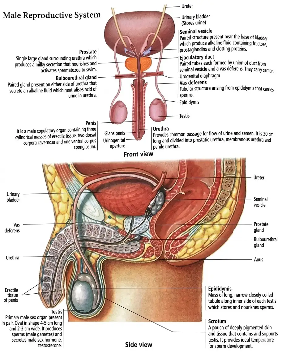
Memory Aids
- Cryptorchidism = Undescended testis.
- Inguinal hernia = Intestine in scrotum.
Quick Checkpoints
- What causes cryptorchidism?
- What is inguinal hernia?
Gametogenesis
Overview
- Primary sex organs (testis in males, ovaries in females) produce gametes (sperms and ova).
- Process of formation of male and female gametes is called gametogenesis.
Memory Aids
- Gametogenesis = formation of gametes (sperm + ovum).
Quick Checkpoints
- What organs produce gametes?
- Define gametogenesis.
Spermatogenesis
Phases and Process
- Occurs in seminiferous tubules of testes, starts at puberty.
- Multiplication phase: Spermatogonia divide mitotically to form many spermatogonia.
- Growth phase: Spermatogonium grows into primary spermatocyte (diploid, 46 chromosomes).
- Maturation phase: Primary spermatocyte undergoes meiosis I to form two secondary spermatocytes (haploid, 23 chromosomes each).
- Secondary spermatocytes undergo meiosis II to form spermatids.
- Each primary spermatocyte produces four spermatids (haploid).
Memory Aids
- Spermatogenesis phases: Multiply, Grow, Mature.
- 1 primary spermatocyte → 4 spermatids.
Quick Checkpoints
- Where does spermatogenesis occur?
- What are the phases of spermatogenesis?
- How many spermatids are formed from one primary spermatocyte?
Formation of Spermatozoa
Structure and Process
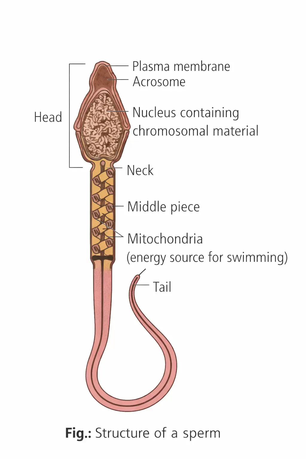
- Transformation of spermatids into spermatozoa is called spermiogenesis or spermateliosis.
- Sperm parts: head, neck, middle piece, tail.
- Head contains haploid nucleus with acrosome (enzymes for fertilisation).
- Middle piece has mitochondria for energy; tail has 9+2 microtubule arrangement for motility.
- About 200-300 million sperms ejaculated per coitus.
- Spermiation: release of spermatozoa from seminiferous tubules.
- Semen = sperm + seminal plasma from accessory glands.
Memory Aids
- Spermiogenesis = spermatids to spermatozoa.
- Sperm structure: Head (nucleus + acrosome), Middle (mitochondria), Tail (movement).
Quick Checkpoints
- What is spermiogenesis?
- Name the parts of a sperm.
- What is spermiation?
Hormonal Control of Male Reproductive System
Hormonal Regulation
- Puberty triggers GnRH secretion from hypothalamus.
- GnRH stimulates anterior pituitary to release LH and FSH.
- LH stimulates Leydig cells to produce testosterone.
- FSH stimulates Sertoli cells to secrete androgen binding protein (ABP) and inhibin.
- Testosterone and inhibin provide negative feedback on LH and FSH secretion.
Memory Aids
- GnRH → LH + FSH → Testosterone + ABP + Inhibin.
- Negative feedback controls hormone levels.
Quick Checkpoints
- What hormones regulate spermatogenesis?
- Role of LH and FSH in male reproduction?
- What feedback controls hormone secretion?
Oogenesis
Process and Stages
- Formation of mature female gamete (ovum).
- Begins during embryonic development with oogonia formation; no new oogonia after birth.
- Oogonia enter prophase I of meiosis and arrest at diplotene stage (primary oocyte).
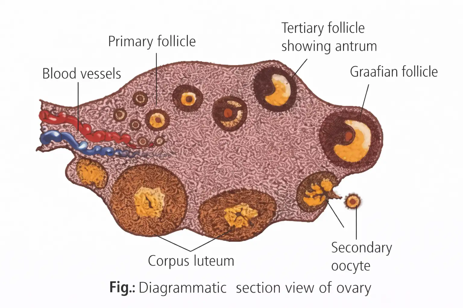
- Primary oocyte surrounded by granulosa cells forms primary follicle.
- Many follicles degenerate; 60,000-80,000 remain at puberty.
- Primary follicles develop into secondary follicles, then tertiary follicles with fluid-filled antrum.
- Primary oocyte completes meiosis I forming large secondary oocyte and small first polar body.
- Secondary oocyte surrounded by zona pellucida; undergoes meiosis II after fertilisation.
- One oogonium produces one ovum and three polar bodies.
- Ovulation: release of secondary oocyte from Graafian follicle.
Memory Aids
- Oogenesis stages: Oogonia → Primary oocyte → Secondary oocyte → Ovum.
- One oogonium → 1 ovum + 3 polar bodies.
- Ovulation releases secondary oocyte.
Quick Checkpoints
- When does oogenesis start?
- What is the fate of primary oocytes before puberty?
- What cells are released during ovulation?
Hormonal Control of Female Reproductive System
Hormonal Regulation
- GnRH from hypothalamus stimulates anterior pituitary to secrete LH and FSH.
- FSH promotes follicle growth and estrogen production; completes meiosis I in oocyte.
- LH stimulates corpus luteum to secrete progesterone.
- High progesterone inhibits GnRH, reducing LH, FSH, and progesterone secretion (negative feedback).
Memory Aids
- GnRH → LH + FSH → Estrogen + Progesterone.
- Progesterone inhibits GnRH and gonadotropins.
Quick Checkpoints
- Which hormones regulate female reproduction?
- Role of FSH and LH in oogenesis?
- How does progesterone regulate hormone levels?
Additional Notes
Key Points
- Vitellogenesis: yolk deposition in primary oocyte during prophase I.
- Mature ovum is spherical, contains nucleus, and is the largest cell in the body.
Memory Aids
- Vitellogenesis = yolk formation in oocyte.
- Ovum = largest cell, spherical with nucleus.
Quick Checkpoints
- What is vitellogenesis?
- Describe the mature ovum.
Menstrual Cycle
Phases and Overview
- Reproductive cycle in female primates (monkeys, apes, humans).
- First menstruation at puberty called menarche.
- Cycle repeats every 28/29 days on average.
- Cycle from one menstruation to next is menstrual cycle.
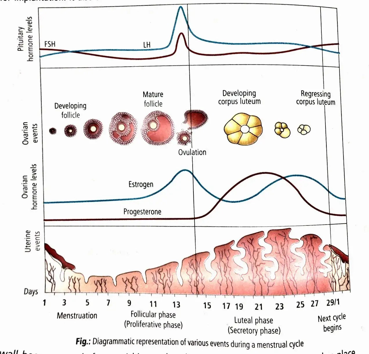
- Four phases: menstrual, follicular, ovulatory, luteal.
Memory Aids
- Menstrual cycle phases: M-F-O-L (Menstrual, Follicular, Ovulatory, Luteal).
- Menarche = first menstruation at puberty.
Quick Checkpoints
- What triggers the start of the menstrual cycle?
- How long is the average menstrual cycle?
- What are the four phases of the menstrual cycle?
Menstrual Phase
Key Features
- Marks cycle start; lasts 3-5 days.
- Day 1 of menstruation = day 1 of cycle.
- LH production drops, corpus luteum degenerates.
- Progesterone and estrogen levels fall rapidly.
- Endometrium breaks down; uterine tissues and blood shed via vagina (menstruation).
- Hygiene during menstruation is crucial.
Memory Aids
- Menstruation = shedding of uterine lining.
- Low LH → corpus luteum degeneration → hormone drop → menstruation.
Quick Checkpoints
- What hormone changes cause menstruation?
- How long does the menstrual phase last?
- What marks day 1 of the menstrual cycle?
Follicular Phase or Proliferative Phase
Key Features
- Occurs days 6-13/14 in 28-day cycle.
- Primary follicles mature into Graafian follicle.
- Gradual rise in LH and FSH secretion.
- FSH stimulates follicle growth and estrogen secretion.
- Estrogen causes endometrium proliferation.
Memory Aids
- Follicle grows → estrogen rises → endometrium thickens.
- FSH and LH increase gradually.
Quick Checkpoints
- Which hormones rise during follicular phase?
- What happens to ovarian follicles in this phase?
- How does estrogen affect the uterus?
Ovulatory Phase
Key Features
- Occurs around day 14 of cycle.
- LH and FSH peak simultaneously.
- LH surge causes rupture of Graafian follicle.
- Ovulation: release of ovum from follicle.
Memory Aids
- LH surge = trigger for ovulation.
- Ovulation occurs mid-cycle (~day 14).
Quick Checkpoints
- What hormone surge triggers ovulation?
- When does ovulation occur in the cycle?
- What is released during ovulation?
Luteal Phase or Secretory Phase
Key Features
- Occurs days 15-28 in 28-day cycle.
- Time between ovulation and next menstruation.
- LH transforms ovulated follicle into corpus luteum.
- Corpus luteum secretes progesterone.
- Progesterone thickens endometrium for implantation.
- Uterine glands produce watery mucus.
- Uterine wall prepared to nourish blastocyst if fertilization occurs.
- No menstruation during pregnancy; cycle stops.
- Without fertilization, corpus luteum degenerates → endometrium breaks down → menstruation starts new cycle.
- Menstrual cycles cease around age 50 (menopause).
- Cyclic menstruation occurs between menarche and menopause.
Memory Aids
- Corpus luteum = progesterone source post-ovulation.
- Progesterone prepares uterus for pregnancy.
- Menopause = end of menstrual cycles (~50 years).
Quick Checkpoints
- What hormone does corpus luteum secrete?
- What happens to the endometrium during luteal phase?
- When does menstruation resume if no fertilization occurs?
Estrous Cycle
Phases and Overview
- Sexual cycle in mammals other than primates (no menstruation).
- Four phases: anestrous, proestrous, estrous, metestrous.
- Anestrous: resting phase, no sexual cycle.
- Proestrous: follicle development, endometrium starts developing under estrogen.
- Estrous: period of heat, Graafian follicle develops under gonadotropins.
- Metestrous: corpus luteum active, produces progesterone; regresses if no pregnancy.
Memory Aids
- Estrous phases: A-P-E-M (Anestrous, Proestrous, Estrous, Metestrous).
- No menstruation in estrous cycle.
Quick Checkpoints
- What is the resting phase in estrous cycle?
- Which phase is called the period of heat?
- What hormone does corpus luteum produce in metestrous?
Fertilisation and Implantation
Process and Site of Fertilisation
- Insemination: Semen released into female vagina during copulation.
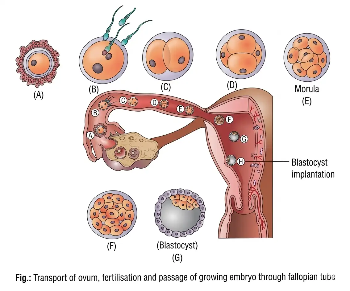
- Secondary oocyte released from mature Graafian follicle enters fallopian tube.
- Ciliated cells in infundibulum propel oocyte to ampulla.
- Ampullary-isthmic junction is fertilisation site.
- Fertilisation requires simultaneous arrival of sperm and ovum at ampulla.
- Sperm aided by uterine and tube contractions, then swim at 1.5-3 mm/min.
Capacitation and Acrosomal Reaction
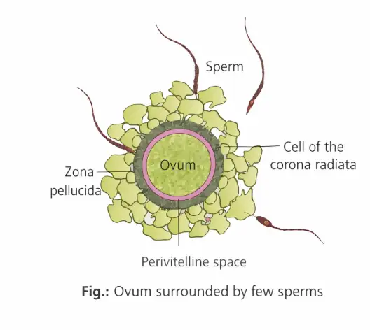
- Capacitation: Activation of sperm by female tract secretions removing surface coatings.
- Exposes sperm receptors; takes 5-6 hours.
- Acrosomal reaction releases lysins: hyaluronidase, corona penetrating enzymes, acrosin.
- Enzymes digest follicle cells, corona radiata, and zona pellucida.
- Plasma membranes of sperm acrosome and oocyte fuse.
- Cortical reaction blocks entry of additional sperms, ensuring monospermy.
Fertilisation and Zygote Formation
- Sperm entry triggers completion of secondary oocyte meiosis II.
- Produces second polar body and haploid ovum (ootid).
- Fusion of haploid sperm and ovum nuclei forms diploid zygote.
- Secondary oocyte viable for fertilisation only 24 hours post-ovulation.
- Fertilizin (egg) and antifertilizin (sperm) interaction causes species-specific sperm adhesion (agglutination).
Sex Determination and Cleavage
- Female gametes carry X chromosome; male gametes carry X or Y (50% each).
- Fusion determines zygote sex: XX female, XY male.
- Zygote undergoes repeated mitotic divisions (cleavage) without volume increase.
- Cleavage forms 2, 4, 8, 16 blastomeres; 8-16 cell stage called morula.
- Morula develops into blastocyst with outer trophoblast and inner cell mass.
- Trophoblast attaches to endometrium; inner cell mass forms embryo.
- Implantation: blastocyst embeds in uterine lining, initiating pregnancy.
Cleavage Significance and Types
- Cleavage distributes cytoplasm among blastomeres.
- Increases protoplasm mobility for cell differentiation and germ layer formation.
- Restores cell size and nucleocytoplasmic ratio.
- Converts unicellular zygote into multicellular embryo.
- Human cleavage is holoblastic (complete division).
- Trophoblast cells near inner cell mass called Rauber cells; form extraembryonic membranes.
- Rauber cells do not form embryo but provide nutrition.
- Inner cell mass is embryo precursor.
Implantation and Zona Hatching
- Implantation occurs ~7 days after fertilisation.
- Zona pellucida prevents premature implantation and exposure of trophoblast cells.
- Blastocyst sheds zona pellucida via zona hatching before implantation.
- Blastocyst inner cell mass faces endometrium for implantation.
- Uterine cells cover blastocyst post-attachment, embedding it in endometrium.
Memory Aids
- Insemination = semen into vagina.
- Capacitation = sperm activation by female secretions.
- Acrosomal reaction = enzyme release to penetrate ovum layers.
- Cortical reaction = blocks polyspermy.
- Cleavage = mitotic division without growth.
- Morula = solid ball; blastocyst = hollow ball.
- Zona hatching = blastocyst escapes zona pellucida for implantation.
- Rauber cells = trophoblast cells near inner cell mass, form membranes.
Quick Checkpoints
- What is the site of fertilisation in the female reproductive tract?
- Define capacitation and its significance.
- Which enzymes are involved in the acrosomal reaction?
- What triggers the completion of meiosis II in the oocyte?
- How is sex determined at fertilisation?
- Describe the stages from zygote to blastocyst.
- What is the role of the trophoblast in implantation?
- Explain the function of zona pellucida during early embryo development.
- What is zona hatching and why is it important?
Pregnancy and Embryonic Development
Implantation and Placenta Formation
- Implantation initiates pregnancy.
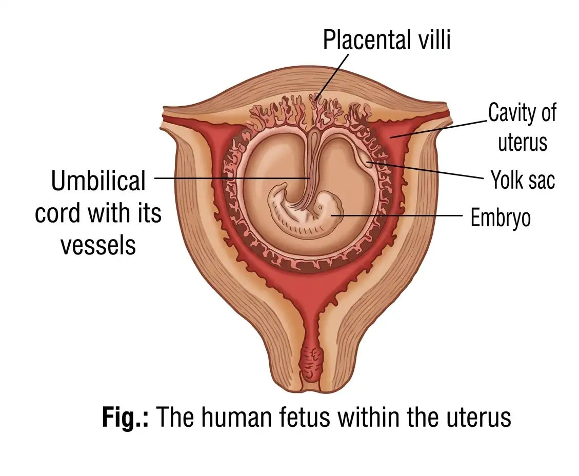
- Trophoblast forms finger-like chorionic villi surrounded by uterine tissue and maternal blood.
- Chorionic villi and uterine tissue interdigitate to form the placenta.
- Placenta supplies oxygen and nutrients, removes wastes, and connects to embryo via umbilical cord.
- Placenta acts as endocrine tissue producing hCG, hPL, estrogens, progestogens, and relaxin (later stage).
- Pregnancy increases maternal blood levels of estrogens, progestogens, cortisol, prolactin, and thyroxine.
Memory Aids
- Placenta = Nutrient + Waste exchange + Hormone production.
- hCG, hPL, relaxin only during pregnancy.
Quick Checkpoints
- What forms the placenta?
- Which hormones are produced by the placenta?
- Role of umbilical cord?
Embryonic Layers and Development
- 8 days post-fertilization, trophoblast splits into syncytiotrophoblast and cytotrophoblast.
- Inner cell mass contains stem cells forming all tissues and organs.
- Inner cell mass differentiates into epiblast and hypoblast forming embryonic disc.
- Gastrulation transforms blastocyst into gastrula with three germ layers: ectoderm, mesoderm, endoderm.
- Epiblast and trophoblast differentiation forms amniotic cavity with amniotic fluid.
- Trophoblast forms extra-embryonic mesoderm, which splits into somatopleuric and splanchnopleuric layers enclosing extraembryonic coelom.
- Chorion formed by somatopleuric mesoderm and trophoblast; main embryonic placenta part and hCG producer.
Memory Aids
- Gastrulation = Formation of 3 germ layers.
- Chorion = Placenta + hCG source.
Quick Checkpoints
- What are the two layers of trophoblast?
- What forms the embryonic disc?
- Function of chorion?
Amnion and Yolk Sac
- Amnion formed by amniogenic cells and somatopleuric membrane.
- Amniotic fluid acts as shock absorber.
- Hypoblast cells line blastocoel forming primary yolk sac (endodermal cells).
- Extraembryonic mesoderm and coelom reduce yolk sac to secondary yolk sac.
- Three germ layers (ectoderm, mesoderm, endoderm) form by cell movements in triploblastic animals.
Memory Aids
- Amniotic fluid = Shock absorber.
- Yolk sac shrinks from primary to secondary.
Quick Checkpoints
- What forms the amnion?
- Role of amniotic fluid?
- Difference between primary and secondary yolk sac?
Gastrulation and Germ Layers
- Primitive streak is a faint groove on epiblast dorsal surface during gastrulation.
- It defines embryo's head-tail and right-left sides.
- Epiblast cells invaginate below primitive streak (invagination).
- Invaginated cells displace hypoblast to form endoderm.
- Cells between epiblast and endoderm form mesoderm.
- Remaining epiblast cells form ectoderm.
- Three germ layers give rise to all tissues and organs with conserved fate across animals.
Memory Aids
- Primitive streak = Body axis + germ layer formation.
- Invagination = Epiblast cells move inward.
- Germ layers: Ectoderm (outer), Mesoderm (middle), Endoderm (inner).
Quick Checkpoints
- What is the primitive streak?
- Which germ layer forms first?
- What is invagination?
Germ Layer Derivatives
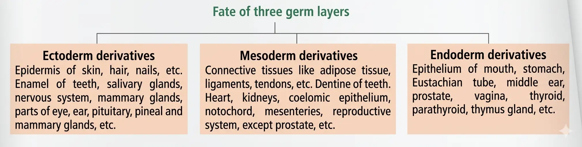
- Ectoderm forms skin, hair, nails, nervous system, parts of eyes and ears.
- Mesoderm forms muscles, bones, connective tissue, heart, kidneys, reproductive organs.
- Endoderm forms lining of mouth, stomach, glands, parts of ear and reproductive system.
- All major body structures arise from these three layers.
Memory Aids
- Ectoderm = External structures + nervous system.
- Mesoderm = Muscles + skeleton + organs.
- Endoderm = Internal linings + glands.
Quick Checkpoints
- Which germ layer forms the nervous system?
- What does mesoderm develop into?
- Endoderm gives rise to which organs?
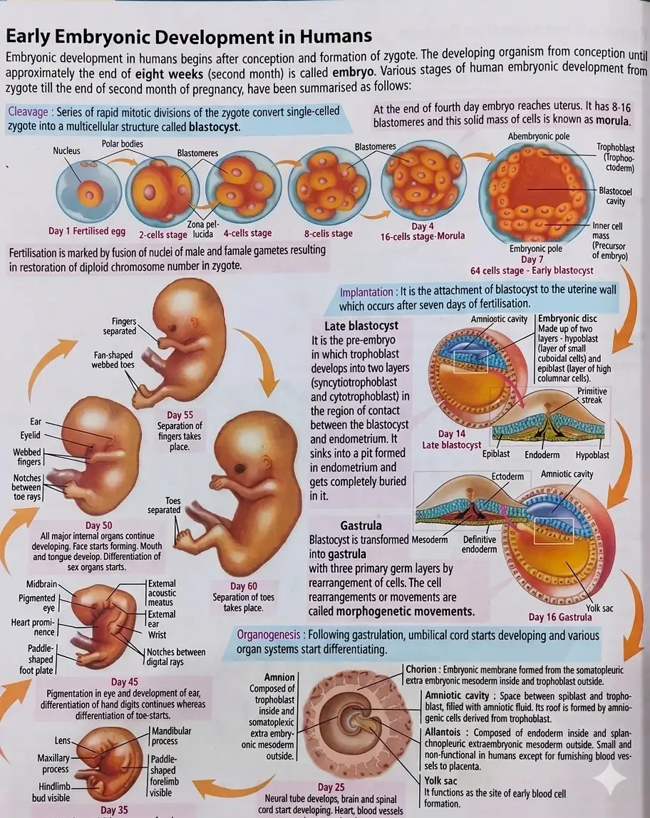
Early Embryonic Development Stages
- Fertilization: Fusion of male and female gametes forming zygote.
- Cleavage: Rapid cell division forming morula (solid ball) by day 4.
- Blastocyst: Hollow structure with inner cell mass and cavity by day 7.
- Implantation: Blastocyst attaches to uterine wall around day 7.
- Late blastocyst: Trophoblast cells develop for implantation.
- Gastrulation (day 16): Formation of three germ layers.
- Organogenesis: Organs develop, umbilical cord forms, systems differentiate.
- Embryo growth (days 35-60): Development of eyes, ears, limbs, fingers, and toes.
- Transforms single cell into complex embryo.
Memory Aids
- Fertilization → Cleavage → Blastocyst → Implantation → Gastrulation → Organogenesis.
- Day 7: Blastocyst and implantation.
- Day 16: Gastrulation starts.
Quick Checkpoints
- What is the sequence of early embryonic stages?
- When does implantation occur?
- What happens during gastrulation?
Parturition and Lactation
Parturition
- Parturition is the expulsion of the full-term baby from the uterus at the end of gestation.
- Signals originate from the fully developed fetus and placenta, causing mild uterine contractions called fetal ejection reflex.
- Fetal ejection reflex triggers oxytocin release from maternal pituitary.
- Oxytocin stimulates strong uterine contractions to expel the baby.
- Relaxin hormone increases flexibility of pubic symphysis and ligaments.
- Relaxin also helps dilate the uterine cervix during labour.
- Forceful uterine contractions during birth are called labour.
- After birth, placenta and umbilical cord remnants are expelled by uterine contractions, called after birth.
Lactation
- Lactation is milk production from mammary glands.
- Mammary glands differentiate during pregnancy and start milk production near term.
- Initial milk produced for 2-3 days is colostrum.
- Colostrum contains antibodies essential for newborn immunity.
Memory Aids
- Fetal ejection reflex = fetus triggers oxytocin release.
- Relaxin = relaxes ligaments and dilates cervix.
- Labour = strong uterine contractions for birth.
- Colostrum = first milk rich in antibodies.
Quick Checkpoints
- What triggers parturition signals?
- Role of oxytocin in birth?
- Function of relaxin during labour?
- What is colostrum and its importance?
- Define after birth.
NEET Zoology — ALL CHAPTERS
1
Structural Organisation in Animals
2
Breathing and Exchange of Gases
3
Body Fluids and Circulation
4
Excretory Products and their Elimination
5
Locomotion and Movement
6
Neural Control and Coordination
7
Human Reproduction
8
Reproductive Health
9
Evolution
10
Microbes in Human Welfare
11
Organism and Population
12
Ecosystem
13
Biodiversity and Conservation