NEET Ug . NEET Zoology . Zoology . Neural Control and Coordination
Chapter 6: Neural Control and Coordination
Ch 6
NEET Zoology
NEET Ug
Introduction
Coordination and Homeostasis
- Coordination is interaction and complementing functions of organs.
- Maintains homeostasis by synchronizing organ functions.
- Example: Physical exercise increases energy demand, oxygen supply, respiration rate, heartbeat, and blood flow.
- Post-exercise, organs like nerves, lungs, heart, kidneys return to normal.
- Neural system and endocrine system jointly coordinate body activities.
- Neural system: fast, point-to-point connections.
- Endocrine system: chemical coordination via hormones.
Memory Aids
- Coordination = organs working together for balance.
- Neural = fast signals; Endocrine = slow hormones.
- Exercise example: increased demand → increased supply.
Quick Checkpoints
- What is coordination in biology?
- How do neural and endocrine systems differ in coordination?
- Why does oxygen supply increase during exercise?
Neural System
Structure and Function
- Composed of specialized cells called neurons.
- Neurons detect, receive, and transmit stimuli.
- Lower invertebrates (e.g., Hydra) have simple neural networks.
- Insects have brains, ganglia, and neural tissues for better organization.
- Vertebrates possess highly developed neural systems.
Memory Aids
- Neurons = nerve cells for signal transmission.
- Hydra = simple nerve net.
- Insects = brain + ganglia; Vertebrates = advanced brain.
Quick Checkpoints
- What are neurons and their role?
- Describe neural organization in Hydra.
- How does neural complexity vary among animals?
Human Neural System
Structure and Divisions
- The human neural system has two parts: central neural system (CNS) and peripheral neural system (PNS).
- CNS includes the brain and spinal cord; it processes information and controls the body.
- PNS consists of all nerves outside the brain and spinal cord connected to CNS.
- PNS nerve fibers are afferent (to CNS) and efferent (from CNS).
- PNS divides into somatic neural system (to skeletal muscles) and autonomic neural system (to involuntary organs and smooth muscles).
- Autonomic system further splits into sympathetic and parasympathetic systems.
- Visceral nervous system is part of PNS controlling impulses between CNS and internal organs.
Memory Aids
- CNS = Brain + Spinal Cord (Control center)
- PNS = Nerves outside CNS (Communication lines)
- Afferent = Arrives at CNS; Efferent = Exits CNS
- Somatic = Skeletal muscles; Autonomic = Involuntary organs
- Sympathetic = Stress response; Parasympathetic = Rest and digest
Quick Checkpoints
- What are the two main parts of the human neural system?
- Which organs form the CNS?
- What types of fibers are in the PNS?
- How is the autonomic system subdivided?
- What is the role of the visceral nervous system?
Peripheral Neural System
Structure and Classification
- Peripheral neural system includes nerves outside brain and spinal cord.
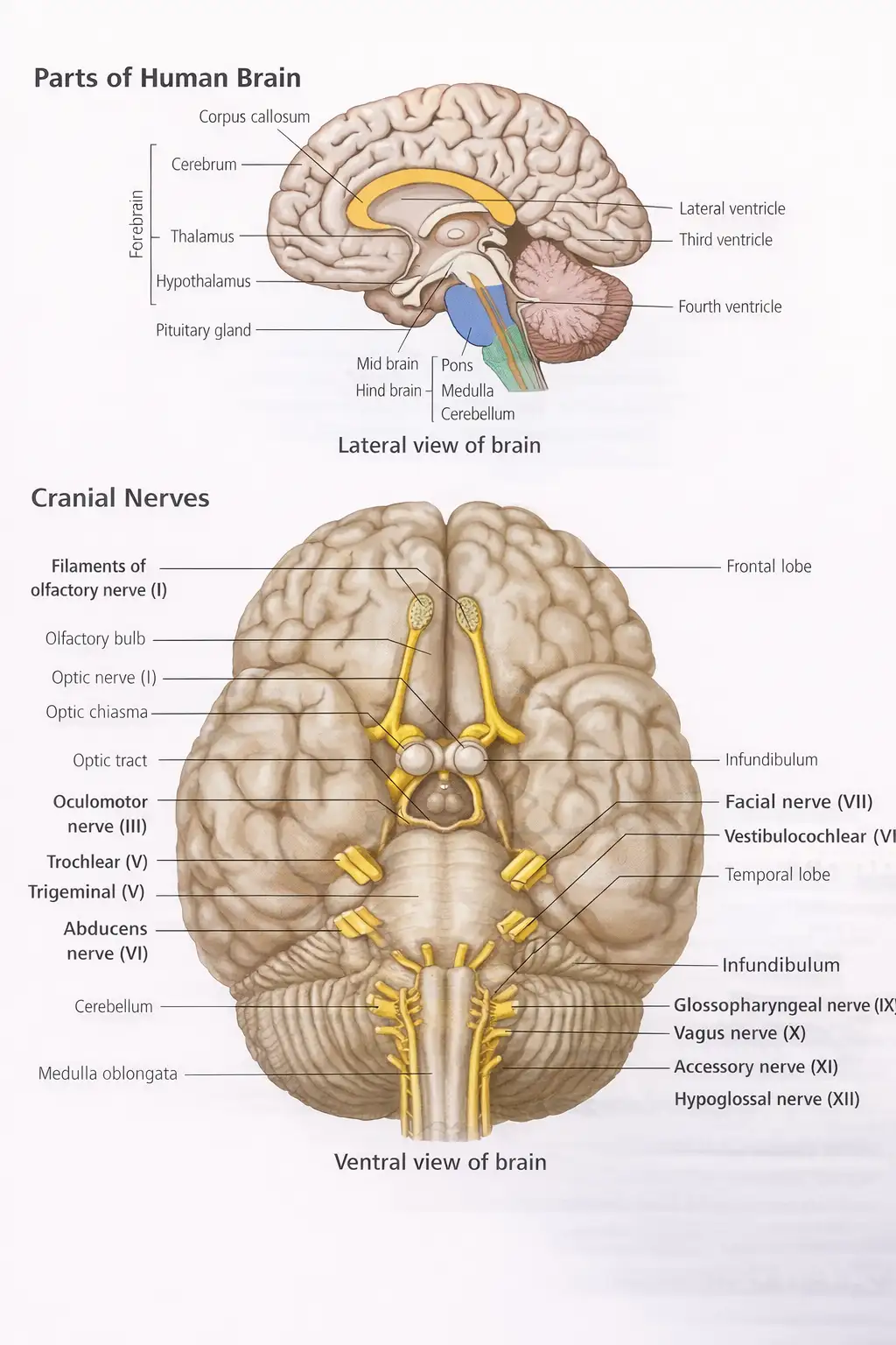
- Two types of nerves: cranial nerves (12 pairs from brain) and spinal nerves (31 pairs from spinal cord).
- Cranial nerves include sensory, motor, and mixed types (e.g., olfactory, optic, facial, vagus).
- Spinal nerves classified into cervical (8 pairs), thoracic (12 pairs), lumbar (5 pairs), sacral (5 pairs), and coccygeal (1 pair).
- PNS subdivides into autonomic neural system and somatic neural system.
- Autonomic system controls involuntary organs; divided into sympathetic (stress response) and parasympathetic (rest) systems.
- Somatic system controls skeletal muscles.
Memory Aids
- Cranial nerves = 12 pairs from brain; Spinal nerves = 31 pairs from spinal cord.
- Autonomic = Involuntary control; Somatic = Voluntary control.
- Sympathetic = Thoracolumbar outflow; Parasympathetic = Craniosacral outflow.
- Sympathetic activates stress responses; Parasympathetic opposes it.
Quick Checkpoints
- What are the two types of peripheral nerves?
- How many pairs of cranial and spinal nerves exist?
- Which nerves are myelinated in PNS?
- What are the functions of sympathetic and parasympathetic systems?
- What does the somatic neural system control?
Neuron as Structural and Functional Unit of Neural System
Structure and Function
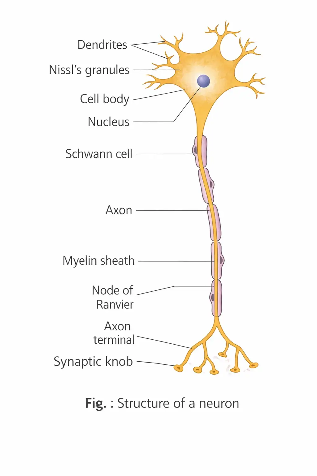
- A neuron has three parts: cell body, dendrites, and axon.
- Cell body contains cytoplasm, organelles, and Nissl's granules (rough ER with ribosomes) for protein synthesis.
- Dendrites are short, branched fibers transmitting impulses toward the cell body; they also contain Nissl's granules.
- Axon is a long fiber with branched distal ends ending in synaptic knobs containing synaptic vesicles with neurotransmitters.
- Axons transmit impulses away from the cell body to synapses or neuromuscular junctions.
Memory Aids
- Neuron parts: Cell body, Dendrites (toward), Axon (away).
- Nissl's granules = protein synthesis sites.
- Synaptic knob = neurotransmitter release point.
Quick Checkpoints
- What are the three main parts of a neuron?
- Where are Nissl's granules found and their function?
- Direction of impulse transmission in dendrites and axons?
Types of Neurons
Classification
- Based on axon and dendrites:
- Unipolar: One axon, found in embryo.
- Bipolar: One axon, one dendrite, found in retina.
- Multipolar: One axon, multiple dendrites, found in cerebral cortex.
- Pseudounipolar: One axon divides into two, e.g., dorsal root ganglion.
- Non-polar: All processes equal, impulse conduction in any direction, e.g., coelenterates.
- Based on function:
- Afferent (sensory): Carry impulses from receptors to CNS; dendrites form receptors.
- Efferent (motor): Carry impulses from CNS to effectors; axon terminals form neuromuscular junction.
Memory Aids
- Unipolar = embryo, Bipolar = retina, Multipolar = brain.
- Afferent = arrive CNS, Efferent = exit CNS.
Quick Checkpoints
- How are neurons classified by structure?
- What is the function of afferent and efferent neurons?
- Examples of pseudounipolar and non-polar neurons?
Types of Axons
Classification and Features
- Two types: myelinated and non-myelinated.
- Myelinated axons are wrapped by Schwann cells forming myelin sheath with nodes of Ranvier; found in spinal and cranial nerves.
- Non-myelinated axons are enclosed by Schwann cells without myelin sheath; found in autonomic and somatic systems.
Memory Aids
- Myelinated = fast, nodes of Ranvier aid impulse speed.
- Non-myelinated = slow, no myelin sheath.
Quick Checkpoints
- What is the role of Schwann cells?
- Where are myelinated and non-myelinated axons found?
- What are nodes of Ranvier?
Generation and Conduction of Nerve Impulse
Resting State and Ionic Gradients
- Neurons are excitable due to polarized membranes with selective ion channels.
- At rest, membrane is more permeable to K+, less to Na+; inside axon has high K+ and negative proteins, outside has high Na+.
- Sodium-potassium pump maintains gradients by pumping 3 Na+ out and 2 K+ in.
- Resting potential is about -70 mV (inside negative, outside positive).
Action Potential and Impulse Conduction
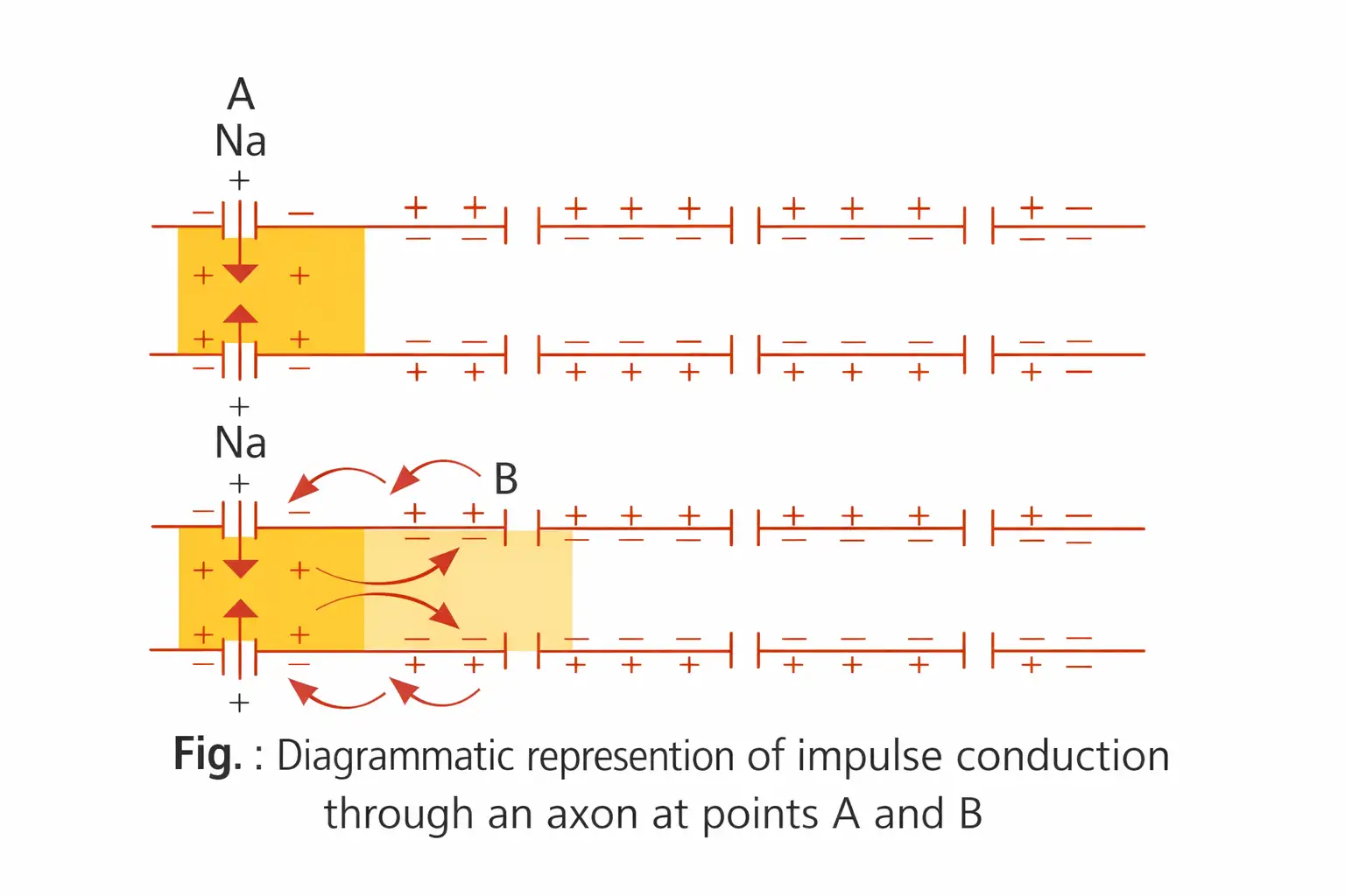
- Stimulus opens Na+ channels, Na+ influx reverses polarity (depolarization) creating action potential (+30 to +45 mV).
- Current flows to adjacent membrane, triggering action potential there; impulse moves along axon.
- After Na+ influx, K+ channels open, K+ efflux restores resting potential (repolarization).
- Conduction is unidirectional; refractory period prevents backward impulse.
- In myelinated axons, impulse jumps node to node (saltatory conduction), faster than continuous conduction in non-myelinated axons.
Memory Aids
- Resting potential = -70 mV, action potential = +30 to +45 mV.
- Na+ in = depolarization, K+ out = repolarization.
- Saltatory conduction = jump nodes, faster impulse.
Quick Checkpoints
- What ions are involved in resting and action potentials?
- How does the sodium-potassium pump maintain resting potential?
- What is saltatory conduction?
Transmission of Impulses
Synapses and Types
- Impulse transmission occurs at synapses between neurons.
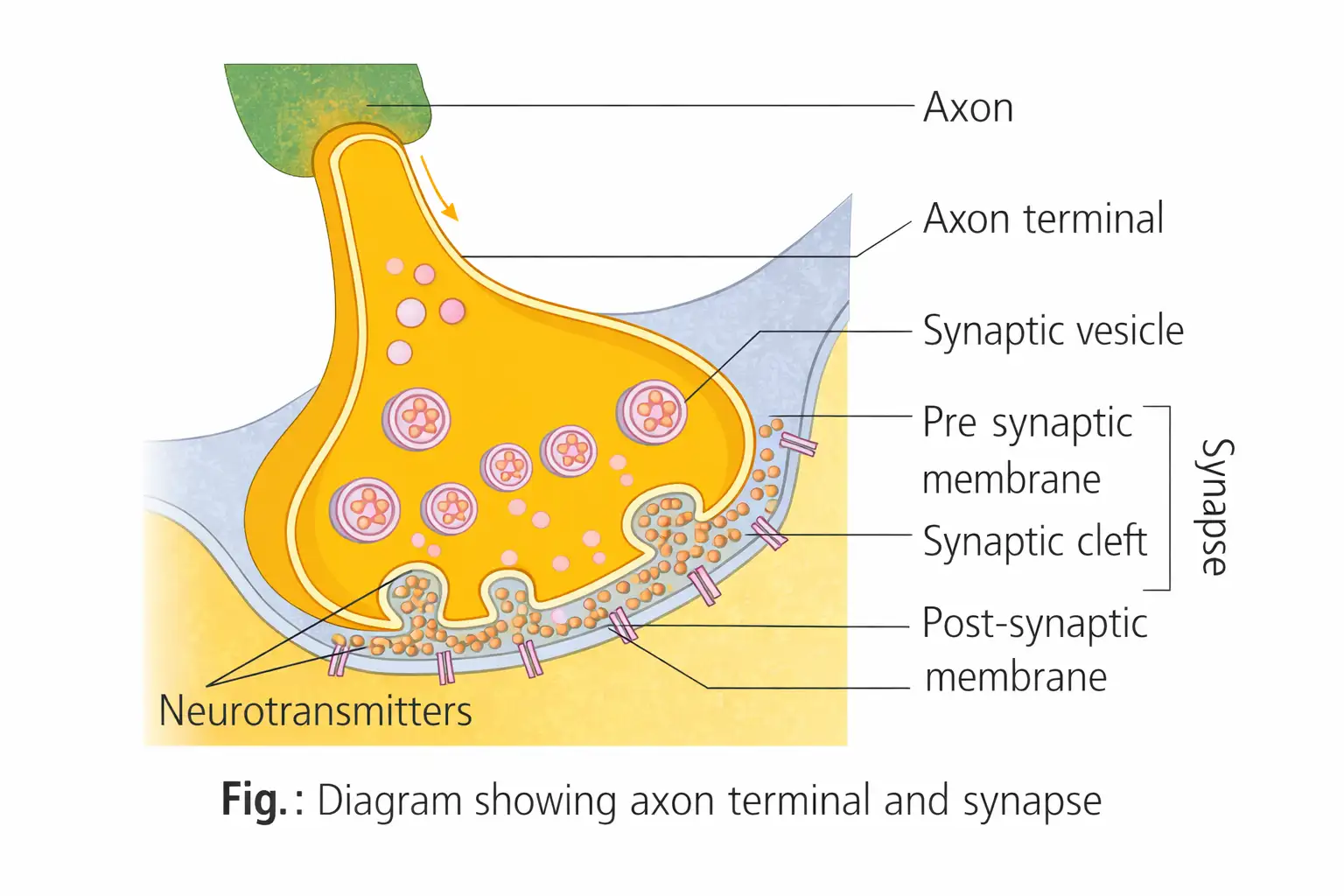
- Synapse = junction of pre-synaptic and post-synaptic neurons, may have synaptic cleft.
- Two types: electrical (direct current flow, rare) and chemical (neurotransmitter-mediated).
Chemical Synapse Mechanism
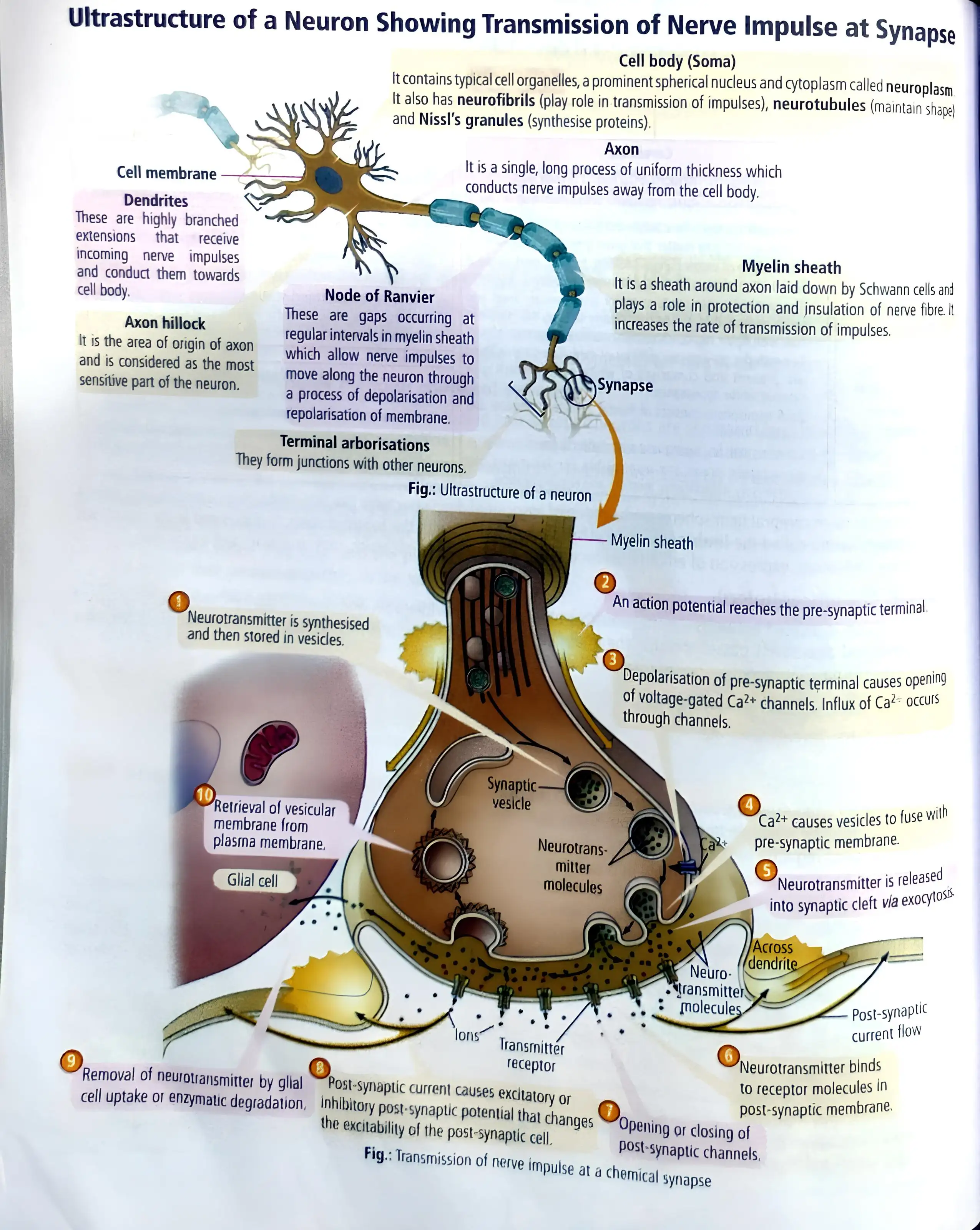
- Axon terminals contain vesicles with neurotransmitters (excitatory: acetylcholine; inhibitory: GABA).
- Impulse arrival causes vesicles to fuse with membrane, releasing neurotransmitters into synaptic cleft.
- Neurotransmitters bind receptors on post-synaptic membrane, opening ion channels to generate new potential.
- New potential can be excitatory or inhibitory.
- Impulse transmission is slower than electrical synapses.
Memory Aids
- Synapse = signal transfer point.
- Neurotransmitters: acetylcholine (excitatory), GABA (inhibitory).
- Chemical synapse slower than electrical.
Quick Checkpoints
- What are the two types of synapses?
- How do neurotransmitters transmit impulses?
- Difference between electrical and chemical synapses?
Central Neural System
Overview
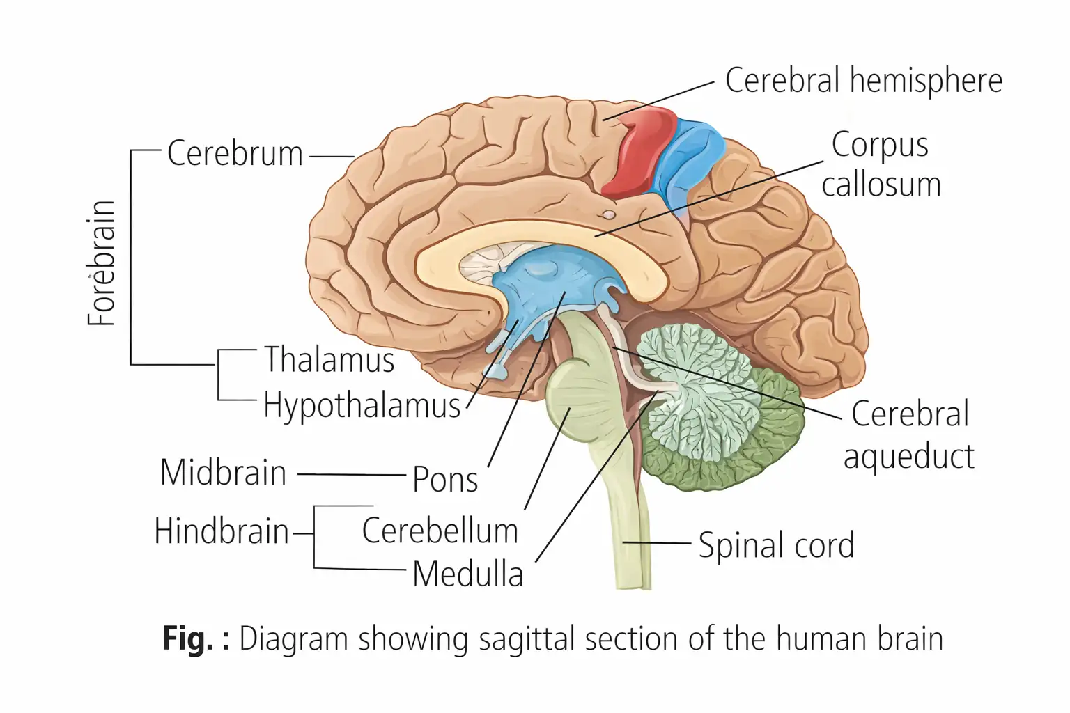
- The brain is the central processing organ, controlling voluntary movements, balance, vital involuntary organs, thermoregulation, hunger, thirst, circadian rhythms, endocrine activities, and behavior.
- Processes vision, hearing, speech, memory, intelligence, emotions, and thoughts.
- Protected by the skull and covered by cranial meninges: dura mater (outer), arachnoid (middle), pia mater (inner).
- Divided into forebrain, midbrain, and hindbrain.
- Cerebrospinal fluid (CSF) cushions the brain, present in ventricles and subarachnoid space.
Memory Aids
- Brain parts: Forebrain (thinking), Midbrain (relay), Hindbrain (balance & vital functions).
- Meninx layers: Dura (durable), Arachnoid (spider-like), Pia (delicate).
Quick Checkpoints
- What are the three main parts of the brain?
- Which meninges layer is the thinnest?
- What is the role of cerebrospinal fluid?
Brain
Structure and Functions
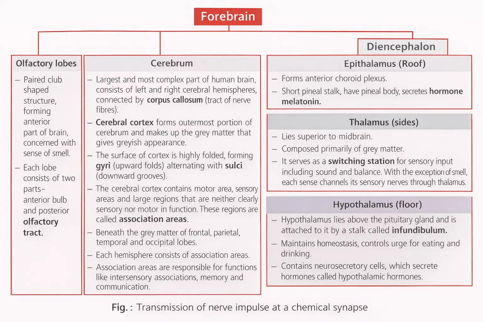
- Forebrain includes olfactory lobes, cerebrum, and diencephalon.
- Cerebrum: largest part, two hemispheres connected by corpus callosum, cerebral cortex with gyri and sulci.
- Diencephalon: epithalamus (melatonin secretion), thalamus (sensory relay except smell), hypothalamus (controls hunger, thirst, hormones).
- Limbic system (amygdala, hippocampus) regulates emotions, sexual behavior, motivation.
- Midbrain: between forebrain and hindbrain, contains cerebral aqueduct and corpora quadrigemina (superior colliculi for sight, inferior colliculi for hearing).
- Hindbrain: pons (fiber tracts, breathing regulation), cerebellum (movement coordination, balance), medulla oblongata (controls respiration, cardiovascular reflexes).
- Brain stem (midbrain, pons, medulla) connects brain to spinal cord, entry/exit for 10 cranial nerves.
- Spinal cord: extends from medulla to second lumbar vertebra, conducts impulses, controls reflexes, has grey (inner) and white (outer) matter, ends as conus medullaris with filum terminale.
Memory Aids
- Forebrain = Olfactory + Cerebrum + Diencephalon.
- Corpora quadrigemina: Superior = Sight, Inferior = Hearing.
- Brain stem = Midbrain + Pons + Medulla.
- Limbic system controls emotions and motivation.
Quick Checkpoints
- What are the main parts of the forebrain?
- Which part of the brain controls balance and coordination?
- What is the function of the thalamus?
- Where is the cerebral aqueduct located?
- What does the medulla oblongata regulate?
- What connects the two cerebral hemispheres?
- What is the role of the spinal cord?
NEET Zoology — ALL CHAPTERS
1
Structural Organisation in Animals
2
Breathing and Exchange of Gases
3
Body Fluids and Circulation
4
Excretory Products and their Elimination
5
Locomotion and Movement
6
Neural Control and Coordination
7
Human Reproduction
8
Reproductive Health
9
Evolution
10
Microbes in Human Welfare
11
Organism and Population
12
Ecosystem
13
Biodiversity and Conservation