NEET Ug . NEET Zoology . Zoology . Locomotion and Movement
Chapter 5: Locomotion and Movement
Ch 5
NEET Zoology
NEET Ug
Introduction
Overview of Movement
- Movement is a basic feature of living beings.
- Animals and plants show various movements; unicellular organisms like Amoeba move by protoplasmic streaming.
- Movement types include cilia, flagella, and tentacles; humans move limbs, jaws, eyelids, tongue, etc.
- Locomotion is voluntary movement causing change in place: walking, running, climbing, flying, swimming.
- All locomotion is movement, but not all movement is locomotion.
- Examples: Paramecium uses cilia for food movement and locomotion; Hydra uses tentacles for prey capture and locomotion; humans use limbs for posture and locomotion.
- Locomotion helps in searching food, shelter, mates, breeding grounds, favorable climate, or escaping predators.
Memory Aids
- Locomotion = movement with change in place.
- Amoeba moves by streaming; Paramecium and Hydra use cilia/tentacles.
- Humans move limbs for posture and locomotion.
Quick Checkpoints
- What distinguishes locomotion from general movement?
- How do unicellular organisms move?
- Examples of locomotion in different organisms?
- Purpose of locomotion in animals?
Types of Movement
Cellular Movement Types
- Amoeboid/Pseudopodial movement: Movement by pseudopodia via cytoplasmic streaming; seen in Amoeba, macrophages, leucocytes; involves microfilaments.
- Ciliary movement: Movement by cilia in internal organs; clears microbes/dust in respiratory tract; transports ova in fallopian tubes and sperm in vasa efferentia.
- Muscular movement: Movement of limbs, jaws, tongue by alternate muscle contraction and relaxation.
- Flagellar movement: Movement by tail and middle piece propulsion; seen in human sperm, sponges' water current, and protozoans like Euglena.
Locomotion requires coordinated muscular, skeletal, and neural activity.
Memory Aids
- Amoeboid = pseudopodia + cytoplasmic streaming.
- Cilia clear and transport in body tubes.
- Muscles contract-relax for movement.
- Flagella propel sperm and protozoans.
Quick Checkpoints
- What causes amoeboid movement?
- Where is ciliary movement found in humans?
- How do muscles produce movement?
- Examples of flagellar movement?
- What systems coordinate locomotion?
Muscle
Overview
- Specialised tissue of mesodermal origin.
- Contributes 40-50% of adult human body weight.
- Properties: excitability, contractility, extensibility, elasticity.
- Three types based on location: Skeletal, Smooth, Cardiac.
Memory Aids
- Muscle = Mesoderm origin + Movement + Multiple types.
- Excitability and contractility are key muscle traits.
Quick Checkpoints
- What is the origin of muscle tissue?
- Name the three types of muscles.
- List special properties of muscles.
Skeletal Muscles
Characteristics
- Associated with skeleton; also called striped or striated muscles.
- Located in limbs, body wall, tongue, pharynx, upper oesophagus.
- Voluntary control by nervous system.
- Functions: locomotion, posture changes.
Memory Aids
- Skeletal = Striated + Voluntary + Attached to bones.
Quick Checkpoints
- Where are skeletal muscles found?
- Are skeletal muscles voluntary or involuntary?
- What actions do skeletal muscles perform?
Cardiac Muscles
Characteristics
- Found in heart walls and large veins entering heart (pulmonary veins, superior vena cava).
- Cylindrical, striated, branched, uninucleate fibers.
- Involuntary control.
- Intercalated discs transmit contraction waves between fibers.
Memory Aids
- Cardiac = Heart muscle + Involuntary + Intercalated discs.
Quick Checkpoints
- Where are cardiac muscles located?
- Describe the structure of cardiac muscle fibers.
- What is the function of intercalated discs?
Smooth Muscles
Characteristics
- Also called visceral, unstriped, or non-striated muscles; spindle-shaped.
- Located in walls of hollow visceral organs: posterior oesophagus, stomach, intestine, lungs, urinary and genital tracts, bladder, blood vessels, iris, dermis.
- Not attached to skeleton.
- Involuntary control by autonomic nervous system.
- Functions: transport of food in digestive tract, movement of gametes in genital tract.
Memory Aids
- Smooth = Spindle shape + Visceral organs + Involuntary.
Quick Checkpoints
- What shape are smooth muscle fibers?
- Where are smooth muscles found?
- Are smooth muscles voluntary or involuntary?
Structure of Skeletal System
Organization
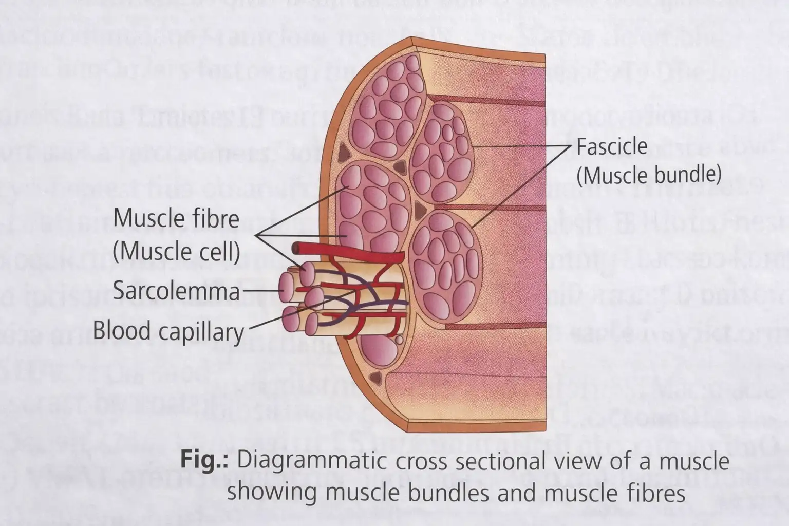
- Muscle made of bundles called fascicles, held by connective tissue called fascia.
- Fascicles contain muscle fibers; fibers are long, cylindrical cells surrounded by sarcolemma.
- Muscle fiber cytoplasm = sarcoplasm, contains many nuclei (syncytium), mitochondria (sarcosomes), and sarcoplasmic reticulum storing calcium ions.
- Muscle fibers contain parallel filaments called myofilaments or myofibrils with alternating dark and light bands.
Striation and Bands
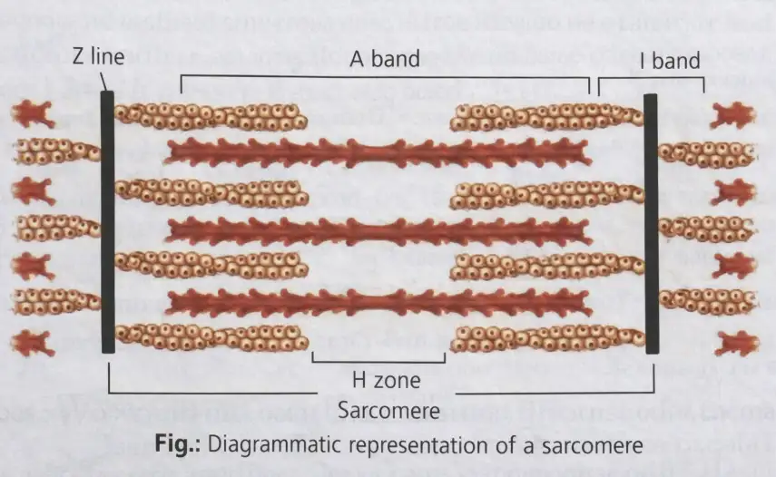
- Striations due to actin (thin) and myosin (thick) protein arrangement.
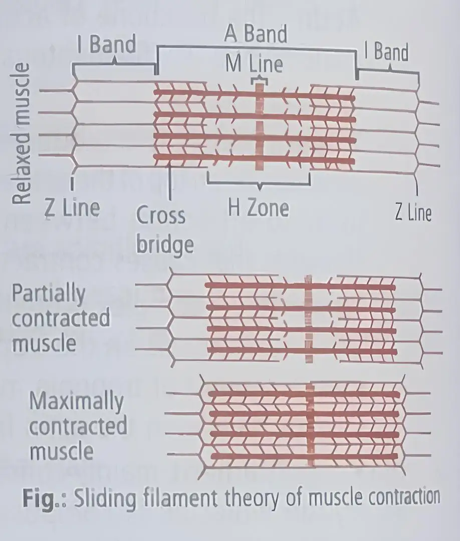
- Light band = 'I'-band (isotropic) with actin; dark band = 'A'-band (anisotropic) with myosin.
- Thin and thick filaments arranged rod-like, parallel to myofibril axis.
- Center of 'I' band has elastic 'Z' line (Z disc) where thin filaments attach.
- Thick filaments held at center of 'A' band by 'M' line.
- Segment between two Z lines = sarcomere, functional contractile unit.
- Resting sarcomere has overlapping thin filaments on thick filaments edges; central thick filament region without overlap = 'H' zone.
Memory Aids
- Striations = Actin (I-band) + Myosin (A-band).
- Sarcomere = Z line to Z line segment.
- H zone = Thick filament center without thin filament overlap.
Quick Checkpoints
- What causes muscle striations?
- Define sarcomere.
- What is the 'H' zone?
Structure of Contractile Proteins
Protein Components
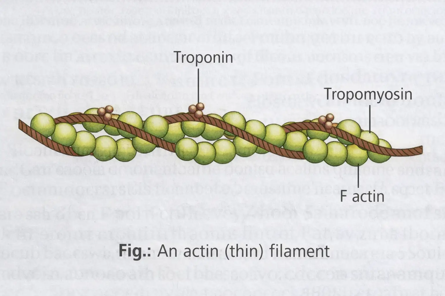
- Four contractile proteins: myosin, actin, troponin, tropomyosin.
- Thin myofilament proteins:
- Actin: Double-stranded F-actin helix made of G-actin monomers.
- Tropomyosin: Double-stranded a-helical rod on F-actin; blocks active sites in resting state.
- Troponin: Complex protein on tropomyosin; masks myosin binding sites on actin in resting state.
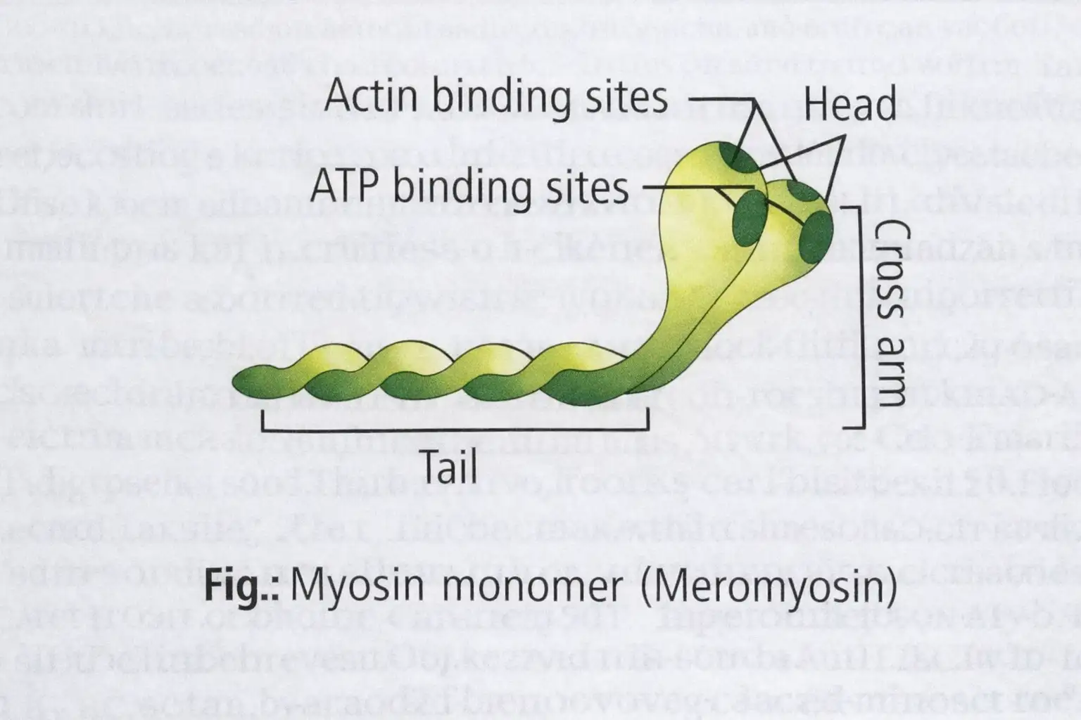
- Thick myofilament mainly myosin:
- Myosin has two heavy and four light polypeptide chains forming two globular heads and a long tail.
- Heads form crossbridges; split by trypsin into heavy meromyosin (HMM) and light meromyosin (LMM).
- HMM heads have ATP and actin binding sites.
Memory Aids
- Thin filament = Actin + Tropomyosin + Troponin.
- Thick filament = Myosin with heads for ATP and actin binding.
Quick Checkpoints
- What proteins form thin myofilaments?
- Describe the structure of myosin.
- What is the role of troponin?
Mechanism of Muscle Contraction
Sliding Filament Theory
- Contraction by sliding of thin filaments over thick filaments.
- Proposed by A. F. Huxley & Ralph Niedergerke, and H.E. Huxley & Jean Hanson (1954).
- Initiated by CNS signal via motor neuron; motor unit = motor neuron + connected muscle fibers.
- Neuromuscular junction (motor end plate) releases acetylcholine, generating action potential in sarcolemma.
- Action potential causes Ca2+ release into sarcoplasm.
- Ca2+ binds troponin, exposing myosin binding sites on actin.
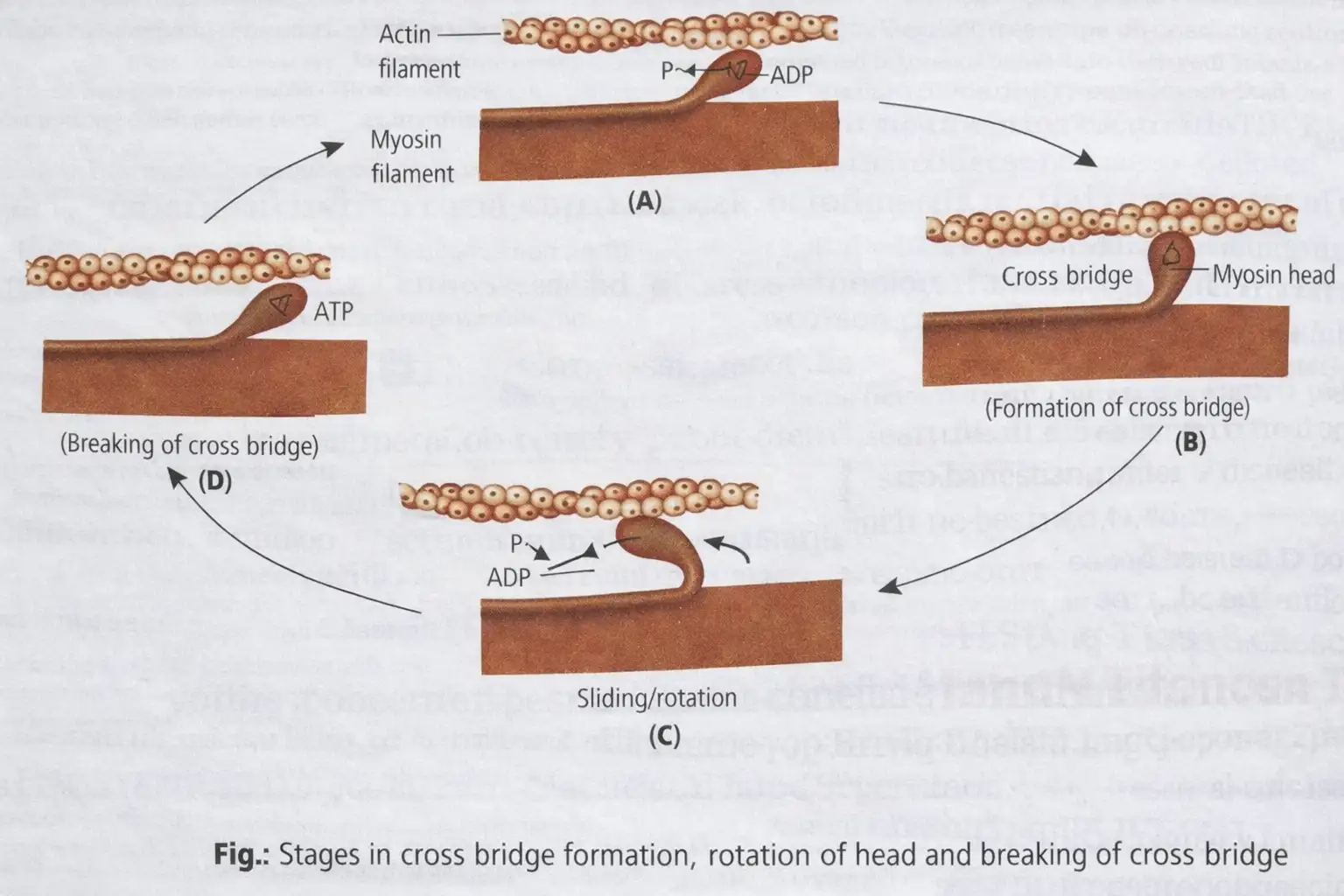
- Myosin heads bind actin using ATP energy, forming cross bridges.
- Myosin heads pivot, pulling actin towards sarcomere center, shortening sarcomere (contraction).
- 'I' bands shorten; 'A' bands remain constant length.
- ADP and phosphate released; ATP binds myosin head, breaking cross bridge.
- ATP hydrolyzed; cycle repeats until Ca2+ pumped back, masking actin sites (relaxation).
- Repeated contraction causes lactic acid buildup, leading to fatigue.
Memory Aids
- Sliding filament = Thin filaments slide over thick filaments.
- Ca2+ triggers exposure of myosin binding sites.
- ATP powers cross bridge cycling.
Quick Checkpoints
- What triggers muscle contraction at molecular level?
- Describe the role of ATP in contraction.
- What happens to sarcomere bands during contraction?
Additional Muscle Concepts
Key Points
- All-or-none law: muscle fiber contracts fully or not at all.
- Oxygen debt: anaerobic respiration during vigorous exercise produces lactic acid causing fatigue; extra oxygen needed to oxidize lactic acid.
- Rigor mortis: post-death muscle stiffening due to lack of ATP preventing actin-myosin separation.
- Antagonistic muscles: muscles acting in opposition, e.g., biceps (flexor) and triceps (extensor).
Memory Aids
- All-or-none: full contraction or none.
- Oxygen debt = lactic acid buildup + fatigue.
- Rigor mortis = no ATP, muscles stiffen after death.
- Antagonistic muscles = opposite actions.
Quick Checkpoints
- What is the all-or-none law?
- Explain oxygen debt in muscles.
- What causes rigor mortis?
- Define antagonistic muscles with example.
Joints
Structure and Function
- Joints connect bones or bones to cartilage, enabling movement.
- Ligaments connect bones; tendons connect bones to muscles.
- Muscle force acts on joints, which serve as fulcrums for movement.
- Joint mobility varies by structure and type.
- Three main types: fibrous, cartilaginous, synovial.
Types of Joints
- Fibrous joints: No movement; bones joined by dense fibrous tissue. Example: skull sutures, teeth to maxilla/mandible.
- Cartilaginous joints: Bones joined by cartilage; allow limited movement. Example: vertebral joints, pubic symphysis.
- Synovial joints: Have fluid-filled synovial cavity; allow free movement. Examples:
- Ball and socket (humerus and pectoral girdle)
- Hinge (knee joint)
- Pivot (atlas and axis)
- Gliding (carpals)
- Saddle (carpal and metacarpal of thumb)
Memory Aids
- Fibrous = Fixed (no movement)
- Cartilaginous = Cartilage connects, allows some movement
- Synovial = Synovial fluid allows free movement
- Examples: Ball & Socket = Shoulder; Hinge = Knee; Pivot = Neck; Gliding = Wrist; Saddle = Thumb
Quick Checkpoints
- What connects bones and muscles?
- Which joint type allows no movement?
- Where is synovial fluid found?
- Name a joint that permits limited movement.
- Identify examples of synovial joints.
Disorders of Muscular and Skeletal System
Overview
- Includes diseases affecting bones and muscles.
- Common disorders involve mineral loss, genetic mutations, autoimmune issues, and metabolic defects.
Memory Aids
- Disorders = Bone + Muscle problems.
- Focus on mineral loss, gene mutation, immunity, metabolism.
Quick Checkpoints
- What causes mineral loss in bones?
- How do gene mutations affect muscles?
- What is the role of immunity in muscle disorders?
Osteoporosis
Key Points
- Age-related bone disease with loss of minerals and fibers from bone matrix.
- Major causes: hormone imbalances (calcitonin, parathormone, estrogen) and deficiencies of calcium and vitamin D.
Memory Aids
- Osteo = bone, porosis = porous (weak bones).
- Think: Hormone + mineral deficiency = weak bones.
Quick Checkpoints
- Which hormones regulate bone minerals?
- How does vitamin D deficiency affect bones?
Muscular Dystrophy
Key Points
- Caused by mutated gene on X-chromosome.
- Mutation leads to absence of dystrophin protein in skeletal muscles.
- Dystrophin deficiency prevents calcium release from muscle cells, blocking muscle contraction.
Memory Aids
- Muscular Dystrophy = X-linked gene mutation.
- Dystrophin missing = no muscle contraction.
Quick Checkpoints
- What is the role of dystrophin?
- Why does muscle contraction fail?
Myasthenia Gravis
Key Points
- Autoimmune disorder affecting neuromuscular junction.
- Leads to fatigue, weakening, and paralysis of skeletal muscles.
Memory Aids
- Myasthenia = muscle weakness.
- Autoimmune attack blocks nerve-muscle communication.
Quick Checkpoints
- What part of muscle function is affected?
- How does immunity cause muscle weakness?
Osteomalacia or Rickets
Key Points
- Occurs in childhood (rickets) or adults (osteomalacia).
- Bones have insufficient calcium and phosphorus.
Memory Aids
- Rickets = childhood bone softening.
- Low calcium/phosphorus = weak bones.
Quick Checkpoints
- What minerals are deficient?
- How does deficiency affect bone strength?
Arthritis
Key Points
- Inflammation of joints.
- Types: rheumatoid arthritis, osteoarthritis, infectious arthritis.
Memory Aids
- Arthritis = joint inflammation.
- Remember types by cause: autoimmune, wear & tear, infection.
Quick Checkpoints
- What causes joint inflammation?
- How do types differ?
Gout
Key Points
- Defect in purine metabolism.
- Excess uric acid and salts accumulate in joints.
Memory Aids
- Gout = uric acid buildup.
- Think purine metabolism defect → joint pain.
Quick Checkpoints
- What metabolic defect causes gout?
- Where does uric acid accumulate?
Tetany
Key Points
- Rapid muscle spasms due to low calcium (Ca++) in body fluids.
Memory Aids
- Tetany = muscle spasms from low Ca++.
Quick Checkpoints
- What ion deficiency causes tetany?
- How does low calcium affect muscles?
Human Skeletal System
Key Points
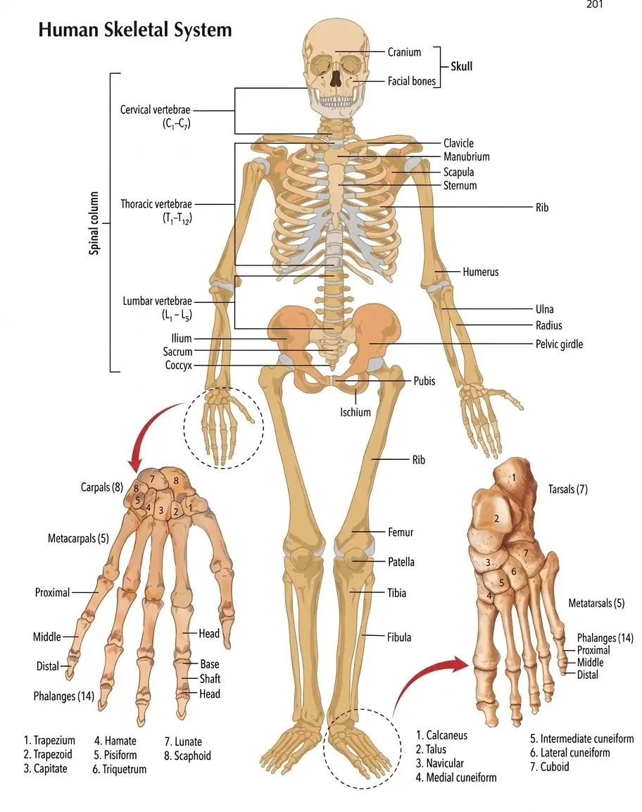
- Divided into skull, spinal column, rib cage, arms, pelvis, and legs.
- Skull: cranium and facial bones protect brain and shape face.
- Spinal column: cervical, thoracic, lumbar vertebrae support body and protect spinal cord.
- Rib cage: ribs, sternum, clavicle, scapula protect chest organs.
- Arms: humerus, radius, ulna bones.
- Pelvic girdle: ilium, pubis, ischium connect spine to legs.
- Legs: femur, patella, tibia, fibula bones.
- Hand bones: carpals, metacarpals, phalanges.
- Foot bones: tarsals, metatarsals, phalanges.
Memory Aids
- Skeleton parts: Head, Spine, Chest, Limbs, Pelvis.
- Hand and foot bones have carpals/tarsals, metacarpals/metatarsals, phalanges.
Quick Checkpoints
- What are the main parts of the skeleton?
- Which bones protect the brain?
- How are hand and foot bones classified?
NEET Zoology — ALL CHAPTERS
1
Structural Organisation in Animals
2
Breathing and Exchange of Gases
3
Body Fluids and Circulation
4
Excretory Products and their Elimination
5
Locomotion and Movement
6
Neural Control and Coordination
7
Human Reproduction
8
Reproductive Health
9
Evolution
10
Microbes in Human Welfare
11
Organism and Population
12
Ecosystem
13
Biodiversity and Conservation