NEET Ug . NEET Zoology . Zoology . Excretory Products and Their Elimination
Chapter 4: Excretory Products and their Elimination
Ch 4
NEET Zoology
NEET Ug
Introduction
Forms of Nitrogenous Wastes
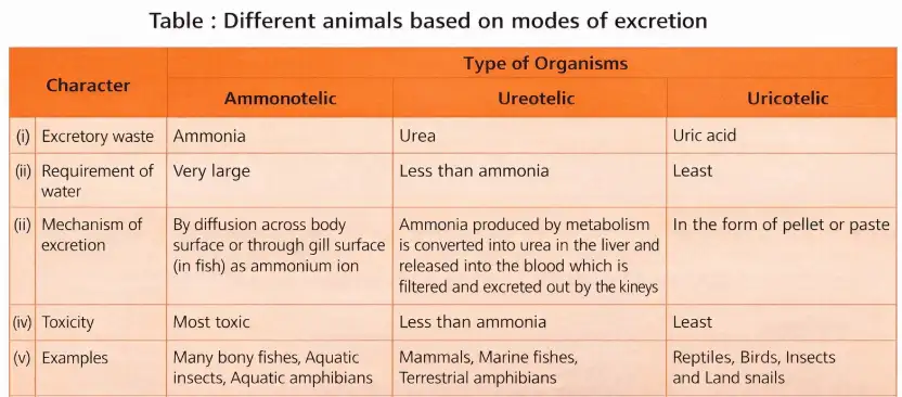
- Excretion removes metabolic wastes like ammonia, urea, uric acid, CO2, water, and ions (Na+, K+, Cl-, phosphate, sulphate).
- Major nitrogenous wastes: ammonia, urea, uric acid.
- Animals differ in waste excretion: ammonotelic (ammonia), ureotelic (urea), uricotelic (uric acid).
Memory Aids
- Ammonotelic = Ammonia excretion, aquatic animals, high water need.
- Ureotelic = Urea excretion, mammals, moderate water need.
- Uricotelic = Uric acid excretion, birds/reptiles, low water need.
Quick Checkpoints
- What are the three main nitrogenous wastes?
- Which animals are ammonotelic, ureotelic, and uricotelic?
- How does water requirement vary among these types?
Excretory Organs
Types of Excretory Organs
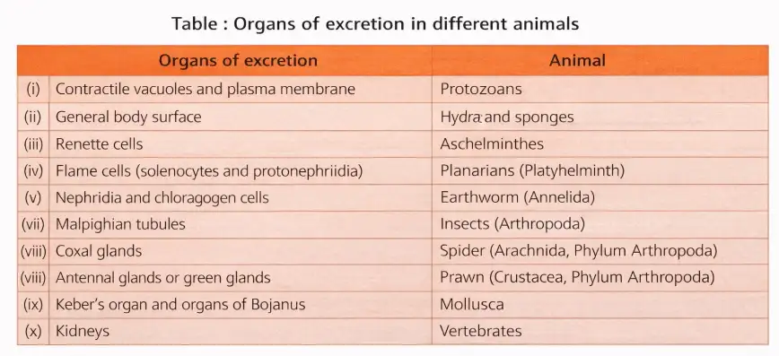
- Protozoans: contractile vacuoles for waste removal.
- Insects: Malpighian tubules.
- Earthworms: nephridia.
- Vertebrates: kidneys.
Memory Aids
- Protozoans = Contractile vacuole.
- Insects = Malpighian tubules.
- Earthworms = Nephridia.
- Vertebrates = Kidneys.
Quick Checkpoints
- Which excretory organ is found in protozoans?
- What organ do insects use for excretion?
- What is the excretory organ in earthworms?
- Which organ do vertebrates use?
Human Excretory System
Components
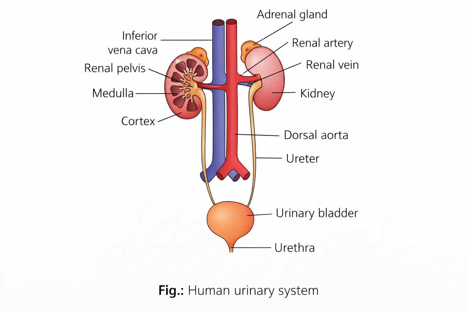
- Pair of kidneys, pair of ureters, urinary bladder, urethra.
Memory Aids
- Kidneys filter blood and produce urine.
- Ureters transport urine to bladder.
- Bladder stores urine.
- Urethra expels urine.
Quick Checkpoints
- What are the main parts of the human excretory system?
- What is the function of ureters?
- Where is urine stored?
- Which tube expels urine?
Kidneys
Structure and Location
- Bean-shaped, reddish brown organs located between last thoracic and third lumbar vertebrae.
- Right kidney is slightly lower than left.
- Size: 10-12 cm length, 5-7 cm width, 2-3 cm thickness; weight: 120-170 g.
- Hilum: notch on inner concave surface for ureter, blood vessels, nerves.
Memory Aids
- Hilum is the entry/exit point for vessels and ureter.
- Right kidney lower due to liver position.
Quick Checkpoints
- Where are kidneys located?
- What is the hilum?
- Which kidney is lower and why?
Kidney Internal Structure
Zones and Parts
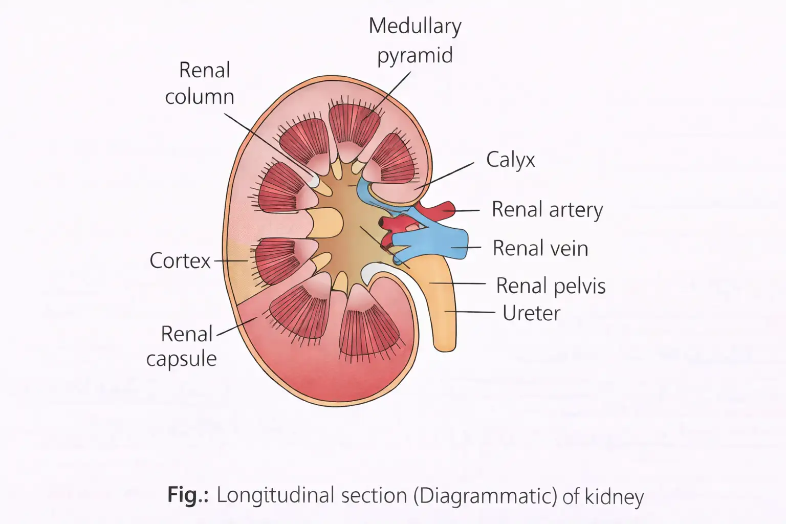
- Outer tough capsule covers kidney.
- Two zones: outer cortex and inner medulla.
- Medulla has medullary pyramids projecting into calyces.
- Cortex extends between pyramids as renal columns (columns of Bertin).
- Major calyces open into renal pelvis leading to ureter.
Memory Aids
- Medullary pyramids collect urine.
- Columns of Bertin separate pyramids.
- Renal pelvis funnels urine to ureter.
Quick Checkpoints
- What are the two main zones of the kidney?
- What are medullary pyramids?
- What is the function of renal pelvis?
Structure of Nephron
Components
- Nephrons are functional units; ~1 million per kidney.
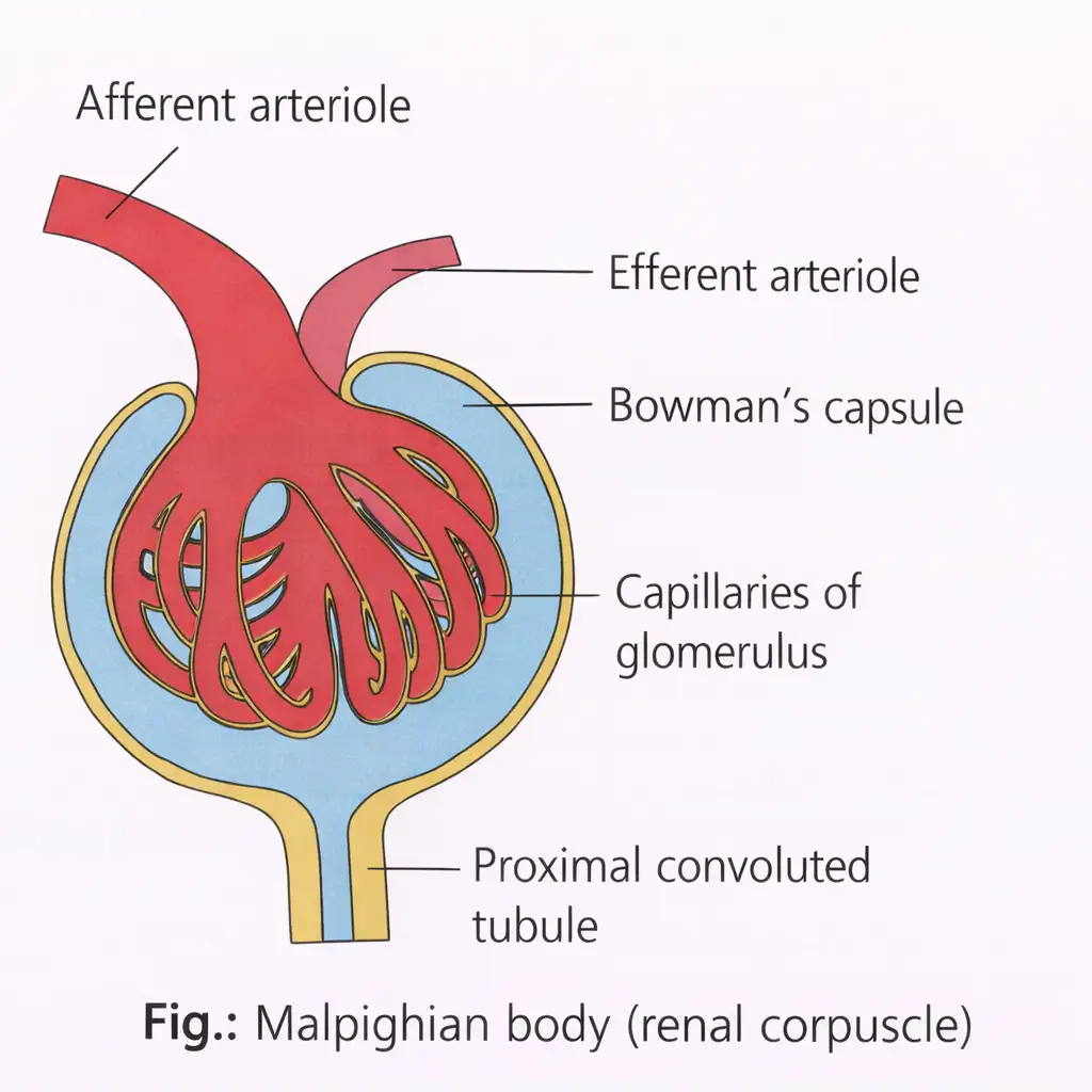
- Two parts: glomerulus (capillary tuft) and renal tubule.
- Renal tubule starts with Bowman's capsule enclosing glomerulus (Malpighian body).
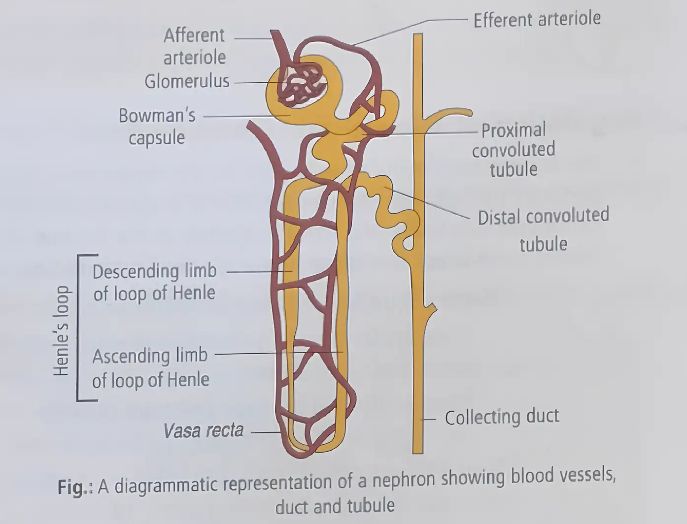
- Renal tubule segments: proximal convoluted tubule (PCT), Henle's loop (descending and ascending limbs), distal convoluted tubule (DCT), collecting ducts.
- Collecting ducts converge into renal pelvis via medullary pyramids.
Memory Aids
- Malpighian body = glomerulus + Bowman's capsule.
- Henle's loop has descending (thin, water permeable) and ascending (thicker, cuboidal cells) limbs.
- Collecting ducts collect urine from many nephrons.
Quick Checkpoints
- What are the two main parts of a nephron?
- What is the Malpighian body?
- Describe the segments of the renal tubule.
- Where does urine collect before entering the renal pelvis?
Types of Kidneys
Classification
- Archinephric kidney: basic ancestral form, in larvae of cyclostomes (Myxine).
- Pronephric kidney: primitive, embryonic stage in some vertebrates.
- Mesonephric kidney: main excretory organ in aquatic vertebrates, temporary in higher vertebrates.
- Metanephric kidney: advanced type with loop of Henle, functional in reptiles, birds, mammals.
Memory Aids
- Archinephric = ancestral.
- Pronephric = embryonic.
- Mesonephric = middle, temporary.
- Metanephric = mature, advanced.
Quick Checkpoints
- What is the ancestral kidney type?
- Which kidney is embryonic?
- Which kidney is temporary in higher vertebrates?
- Which kidney has loop of Henle?
Nephron Zones
Location of Parts
- Malpighian corpuscle, PCT, DCT in renal cortex.
- Loop of Henle, collecting duct, duct of Bellini in renal medulla.
- PCT cells have brush border microvilli for surface area.
- Descending limb: thin, squamous epithelium, permeable to water.
- Ascending limb: thicker, cuboidal epithelium, impermeable to water.
- Efferent arteriole forms peritubular capillaries and vasa recta around tubules.
Memory Aids
- PCT: microvilli increase absorption.
- Descending limb: water permeable.
- Ascending limb: water impermeable.
- Vasa recta supports medullary function.
Quick Checkpoints
- Where are Malpighian corpuscle and PCT located?
- What epithelium lines the descending limb?
- What is the role of vasa recta?
Types of Nephrons
Characteristics
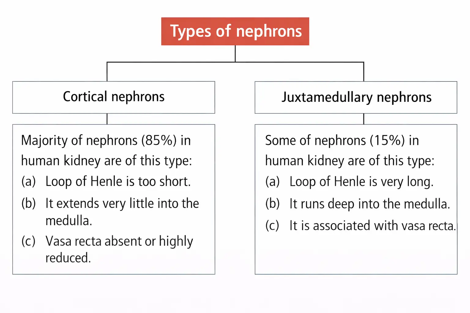
- Cortical nephrons (85%): short loop of Henle, extend little into medulla, few/no vasa recta.
- Juxtamedullary nephrons (15%): long loop of Henle, extend deep into medulla, associated with vasa recta.
Memory Aids
- Cortical = short loop, mostly cortex.
- Juxtamedullary = long loop, deep medulla.
Quick Checkpoints
- What percentage of nephrons are cortical?
- Which nephron type has a long loop of Henle?
- Which nephron type is associated with vasa recta?
Urine Formation
Processes
- Glomerular filtration (ultrafiltration) of blood plasma by glomeruli.
- Tubular reabsorption of useful substances back to blood.
- Tubular secretion of wastes and excess ions into filtrate.
Memory Aids
- Filtration: blood plasma filtered in glomerulus.
- Reabsorption: nutrients and water reabsorbed.
- Secretion: wastes secreted into tubule.
Quick Checkpoints
- What are the three main steps in urine formation?
- Where does filtration occur?
- What is reabsorbed during tubular reabsorption?
- What substances are secreted during tubular secretion?
Glomerular Filtration
Mechanism
- Blood filtered by glomerulus through endothelium, basement membrane, and Bowman's capsule epithelium.
- Podocytes form filtration slits allowing plasma except proteins to pass.
- Filtrate formed is called ultrafiltrate.
- Glomerular filtration rate (GFR) ~125 mL/min (180 L/day).
- Filtration driven by blood hydrostatic pressure; no energy required.
Memory Aids
- Filtration layers: endothelium, basement membrane, podocytes.
- Filtrate excludes proteins.
- GFR is a measure of kidney filtration efficiency.
Quick Checkpoints
- What are the three layers involved in filtration?
- What cells form filtration slits?
- What is the normal GFR value?
Regulation of Glomerular Filtration Rate
Mechanism
- Juxtaglomerular apparatus (JGA) regulates GFR.
- JGA formed by distal convoluted tubule and afferent arteriole contact.
- Drop in GFR triggers renin release from JG cells.
- Renin increases glomerular blood flow, restoring GFR.
Memory Aids
- JGA senses GFR changes.
- Renin release adjusts filtration rate.
Quick Checkpoints
- What is the role of JGA?
- How does renin affect GFR?
Effective Filtration Pressure
Components
- Glomerular blood hydrostatic pressure (GBHP): ~60 mmHg, promotes filtration.
- Blood colloidal osmotic pressure (BCOP): ~32 mmHg, opposes filtration.
- Capsular hydrostatic pressure (CHP): ~18 mmHg, opposes filtration.
- Effective filtration pressure (EFP) = GBHP - (BCOP + CHP) = 10 mmHg.
- EFP drives plasma filtration into Bowman's capsule.
- Renal plasma flow (RPF): 650-700 mL/min out of 1100-1200 mL blood/min.
Memory Aids
- EFP is net pressure for filtration.
- GBHP is main driving force.
- BCOP and CHP resist filtration.
Quick Checkpoints
- What pressures oppose filtration?
- How is EFP calculated?
- What is the typical value of EFP?
Tubular Reabsorption
Process
- Filtrate substances reabsorbed into blood via peritubular capillaries.
- About 99% of filtrate reabsorbed.
- Active reabsorption: glucose, amino acids, Na+.
- Passive reabsorption: nitrogenous wastes, water.
Memory Aids
- Most filtrate is reabsorbed to prevent loss.
- Active transport for nutrients and ions.
- Water reabsorbed passively.
Quick Checkpoints
- What percentage of filtrate is reabsorbed?
- Which substances are actively reabsorbed?
- How is water reabsorbed?
Tubular Secretion
Process
- Tubular cells secrete H+, K+, ammonia into filtrate.
- Maintains ionic and acid-base balance.
- Removes substances not filtered by glomerulus.
- Secretion via active and passive transport from peritubular capillaries.
- If filtration stops due to low pressure, urine forms by secretion only.
Memory Aids
- Secretion removes excess ions and wastes.
- Important for acid-base balance.
Quick Checkpoints
- Which ions are secreted into filtrate?
- Why is tubular secretion important?
- What happens when filtration stops?
Function of the Tubules
Proximal Convoluted Tubule (PCT)
- PCT lined by simple cuboidal brush border epithelium to increase surface area for reabsorption.
- Reabsorbs nearly all essential nutrients and 70-80% of electrolytes and water.
- Maintains pH and ionic balance by secreting H+, NH3, K+ into filtrate and absorbing HCO3-.
- Glucose and amino acids reabsorbed by active transport; water by osmosis.
- Maximum reabsorption occurs in PCT.
Memory Aids
- PCT = "Primary Catch Tubule" for nutrients and water.
- Brush border = More surface, more absorption.
- Secretes acids and bases to balance pH.
Quick Checkpoints
- What epithelium lines the PCT?
- Which substances are actively reabsorbed in PCT?
- How does PCT maintain pH?
Henle's Loop
- Ascending limb has minimal reabsorption; maintains high osmolarity in medulla.
- Descending limb permeable to water, impermeable to electrolytes; concentrates filtrate.
- Ascending limb impermeable to water; actively/passively transports electrolytes out.
- Filtrate dilutes as electrolytes leave ascending limb to medullary fluid.
Memory Aids
- Descending limb = "Water leaves, salts stay".
- Ascending limb = "Salts leave, water stays".
- Loop creates medullary osmotic gradient.
Quick Checkpoints
- Which limb is permeable to water?
- What happens to filtrate concentration in descending limb?
- How does ascending limb affect filtrate?
Distal Convoluted Tubule (DCT)
Functions
- Conditional reabsorption of Na+ and water.
- Reabsorbs HCO3- and selectively secretes H+, K+, and NH3 to maintain pH and ionic balance.
Memory Aids
- DCT = "Dynamic Control Tubule" for ions and pH.
- Selective secretion keeps blood balanced.
Quick Checkpoints
- What ions are reabsorbed in DCT?
- Which ions are secreted to maintain pH?
Collecting Duct
Functions
- Extends from cortex to medulla; reabsorbs large amounts of water to concentrate urine.
- Allows small amounts of urea to pass into medullary interstitium to maintain osmolarity.
- Maintains pH and ionic balance by selective secretion of H+ and K+.
Memory Aids
- Collecting duct = "Final water adjustment".
- Urea recycling helps osmolarity.
- Secretion controls blood pH and ions.
Quick Checkpoints
- What is the main function of the collecting duct?
- How does urea contribute to urine concentration?
- Which ions are secreted here?
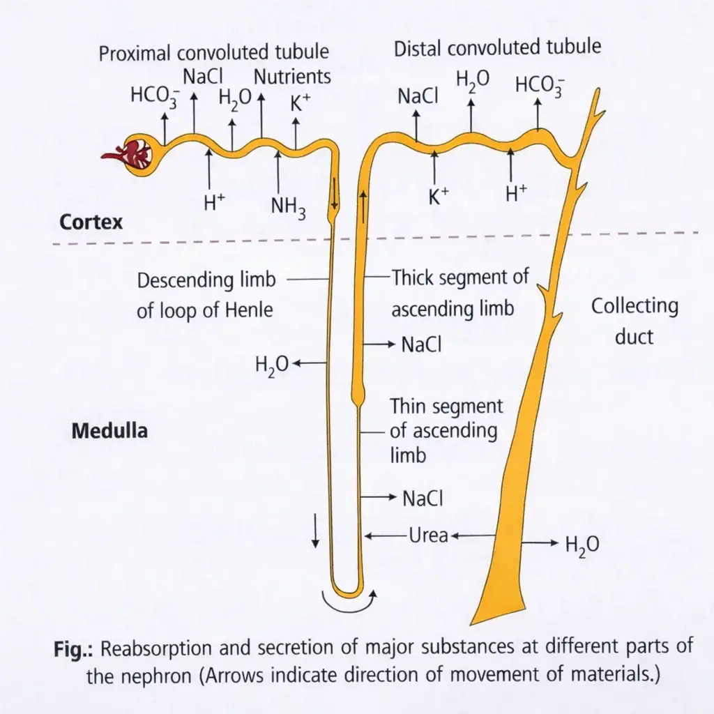
Image Explanation
- Nephron filters blood and removes waste.
- PCT reabsorbs nutrients, water, NaCl, and HCO3-.
- H+ and NH3 secreted into tubule.
- Descending limb reabsorbs water, concentrating filtrate.
- Ascending limb reabsorbs NaCl, diluting filtrate.
- DCT selectively reabsorbs and secretes ions.
- Collecting duct reabsorbs water and urea to concentrate urine.
- Process maintains water, salt, and acid-base balance.
Mechanism of Concentration of the Filtrate
Counter Current System in Kidney
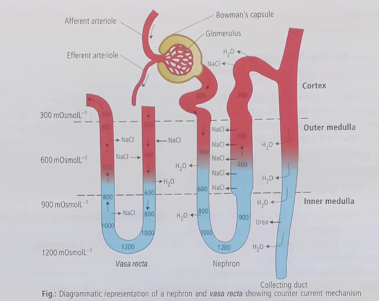
- Mammalian kidneys produce concentrated urine via loop of Henle and vasa recta in the medulla.
- Filtrate flows in opposite directions in the two limbs of loop of Henle, forming a counter current.
- Blood flow in the two limbs of vasa recta is also opposite, creating a counter current pattern.
- This arrangement maintains an osmolarity gradient from cortex (300 mOsmol/L) to inner medulla (1200 mOsmol/L).
- NaCl is actively transported by the ascending limb of Henle's loop and exchanged with descending vasa recta.
- NaCl returns to medullary interstitium via ascending vasa recta.
- Urea enters thin ascending limb and is recycled via collecting tubule, aiding concentration.
- Counter current mechanism conserves water and concentrates urine up to four times the initial filtrate.
- Longer loop of Henle in mammals correlates with higher urine concentration due to more water reabsorption.
Memory Aids
- Counter current = Opposite flows in loop of Henle and vasa recta.
- Osmolarity gradient: Cortex 300 → Inner medulla 1200 mOsmol/L.
- NaCl pumped up ascending limb; water moves down descending limb.
- Urea recycling enhances medullary concentration.
- Longer loop = More water conserved = More concentrated urine.
Quick Checkpoints
- What creates the counter current in the nephron?
- How does NaCl contribute to osmolarity gradient?
- Role of vasa recta in concentration mechanism?
- How does urea aid urine concentration?
- Effect of loop of Henle length on urine concentration?
Regulation of Kidney Function
Hormonal Feedback Mechanism
- Kidney function is regulated by hormones via feedback loops.
- Key regulators: Hypothalamus (ADH), Juxtaglomerular Apparatus (JGA), Heart (ANF).
Memory Aids
- ADH from hypothalamus controls water balance.
- JGA controls blood pressure via renin.
- ANF from heart opposes renin effects.
Quick Checkpoints
- Which hormones regulate kidney function?
- What triggers ADH release?
- How does JGA affect blood pressure?
- Role of ANF in kidney regulation?
Antidiuretic Hormone (ADH)
Function and Regulation
- Osmoreceptors detect changes in blood and body fluid volume and ionic concentration.
- Fluid loss activates osmoreceptors, stimulating hypothalamus to release ADH (vasopressin) from neurohypophysis.
- ADH promotes water reabsorption in distal kidney tubules, reducing urine output (prevents diuresis).
- Increased body fluid volume inhibits ADH release, completing feedback.
- ADH also constricts blood vessels, raising blood pressure and glomerular filtration rate (GFR).
- Diuretics increase urine production, preventing water retention and edema.
- Alcohol inhibits ADH release; caffeine blocks ADH action and sodium reabsorption, increasing urine loss.
- ADH deficiency causes diabetes insipidus with excessive urine output (20-25 L/day).
Memory Aids
- ADH = Anti-Diuresis Hormone.
- Osmoreceptors trigger ADH release on fluid loss.
- Alcohol and caffeine increase urine by affecting ADH.
- Diabetes insipidus = ADH deficiency disease.
Quick Checkpoints
- What activates osmoreceptors?
- How does ADH affect kidney tubules?
- Effect of alcohol and caffeine on ADH?
- What causes diabetes insipidus?
Juxtaglomerular Apparatus (JGA)
Renin-Angiotensin Mechanism
- Decrease in glomerular blood flow/pressure/GFR activates JG cells to release renin.
- Renin converts angiotensinogen to angiotensin I, then to angiotensin II.
- Angiotensin II effects:
- Powerful vasoconstrictor, raises glomerular blood pressure and GFR.
- Stimulates adrenal cortex to release aldosterone.
- Aldosterone promotes sodium and water reabsorption in distal tubules, increasing blood pressure and GFR.
- This is the Renin-Angiotensin mechanism controlling blood pressure and kidney function.
Memory Aids
- Renin released when blood flow drops.
- Angiotensin II = vasoconstrictor + aldosterone stimulator.
- Aldosterone = sodium & water reabsorption.
- Renin-Angiotensin system regulates blood pressure.
Quick Checkpoints
- What triggers renin release?
- Role of angiotensin II?
- How does aldosterone affect kidneys?
- What is the Renin-Angiotensin mechanism?
Atrial Natriuretic Factor (ANF)
Function and Regulation
- Increased blood flow to heart atria triggers ANF release.
- ANF causes vasodilation, lowering blood pressure.
- ANF inhibits renin release from JGA, opposing the renin-angiotensin mechanism.
Memory Aids
- ANF released by atria on high blood flow.
- ANF dilates vessels, reduces blood pressure.
- ANF blocks renin to balance blood pressure.
Quick Checkpoints
- What triggers ANF release?
- How does ANF affect blood vessels?
- What is ANF's role in renin regulation?
Micturition
Mechanism and Process
- Expulsion of urine from the urinary bladder is called micturition.
- Neural control of this process is the micturition reflex.
- Urine formed by nephrons is stored in the urinary bladder.
- Stretching of bladder walls by urine activates stretch receptors.
- Stretch receptors send signals to the central nervous system (CNS).
- CNS sends motor signals to contract bladder smooth muscles.
- Simultaneously, urethral sphincter relaxes to release urine.
- Average urine output in adults is 1 to 1.5 litres per day.
Urine Characteristics
- Urine is light yellow, watery, and slightly acidic (pH ~6.0).
- Colour is due to pigment urochrome from haemoglobin breakdown.
- About 25-30 g of urea is excreted daily.
- Standing urine develops ammonia smell due to bacterial urea breakdown.
- Urine analysis aids diagnosis of metabolic and kidney disorders.
- Glucose (glycosuria) and ketone bodies (ketonuria) indicate diabetes mellitus.
- Albumin presence is albuminuria; WBCs or pus presence is pyuria.
Memory Aids
- Micturition reflex = bladder stretch → CNS signal → muscle contract + sphincter relax.
- Urochrome = urine yellow pigment from RBC breakdown.
- Glycosuria and ketonuria = diabetes markers in urine.
- Albuminuria = albumin in urine; Pyuria = pus/WBCs in urine.
Quick Checkpoints
- What triggers the micturition reflex?
- Which muscles contract and relax during urination?
- What causes the yellow colour of urine?
- How does urine pH and smell change over time?
- What do glycosuria and ketonuria indicate?
- Define albuminuria and pyuria.
Role of Other Organs in Excretion
Lungs
- Remove large amounts of CO2 (200 mL/min).
- Eliminate significant water daily through respiration.
Liver
- Secretes bile containing bilirubin, biliverdin, cholesterol, degraded steroid hormones, vitamins, and drugs.
- These substances exit the body with digestive wastes.
Skin
- Sweat glands produce watery fluid with NaCl, small urea, lactic acid.
- Sweat cools body surface and removes some wastes.
- Sebaceous glands secrete sterols, hydrocarbons, waxes via sebum.
- Sebum protects skin with oily covering.
- Small nitrogenous wastes also eliminated via saliva.
Memory Aids
- Lungs: "CO2 and water out" through breathing.
- Liver: "Bile removes waste pigments and toxins."
- Skin: "Sweat cools and clears; sebum protects."
- Saliva: Minor nitrogenous waste exit.
Quick Checkpoints
- What wastes do lungs primarily remove?
- Which substances does liver secrete in bile?
- What is the main function of sweat besides waste removal?
- What wastes are eliminated by sebaceous glands?
- Can saliva remove nitrogenous wastes?
Uremia
Explanation
- Malfunction of kidneys causes urea accumulation in blood.
- Leads to kidney failure if untreated.
- Hemodialysis removes urea from blood externally.
- Blood is taken from artery, anticoagulant (heparin) added.
- Blood passes through cellophane tube in dialysing fluid without nitrogenous wastes.
- Urea and wastes diffuse out by concentration gradient.
- Cleaned blood returned to body with anti-heparin added.
Memory Aids
- Hemodialysis = "Blood cleaning outside body"
- Heparin prevents clotting during dialysis.
Quick Checkpoints
- What causes uremia?
- How does hemodialysis work?
- Why is dialysing fluid free of nitrogenous wastes?
Kidney Transplantation
Explanation
- Used to treat acute renal failure.
- Transplant functioning kidney from compatible donor.
- Close relatives preferred to reduce rejection risk.
- Immune system may reject incompatible kidneys.
Memory Aids
- Transplant = "Replace failed kidney"
- Compatibility reduces rejection.
Quick Checkpoints
- When is kidney transplantation used?
- Why choose a close relative as donor?
- What causes rejection?
Renal Calculi
Explanation
- Stones formed by crystallized salts (oxalates) in kidney.
- Caused by dehydration, excess uric acid, high milk intake.
- Also due to metabolic imbalance, infection, hormonal issues.
Memory Aids
- Calculi = "Kidney stones"
- Dehydration and diet affect stone formation.
Quick Checkpoints
- What are renal calculi made of?
- List causes of renal calculi.
- How does dehydration contribute?
Glomerulonephritis
Explanation
- Inflammation of kidney's glomerulus.
- Impairs filtration function.
Memory Aids
- Glomerulonephritis = "Glomerulus inflammation"
Quick Checkpoints
- What part of kidney is inflamed?
- How does inflammation affect kidney function?
Human Excretory System
Explanation
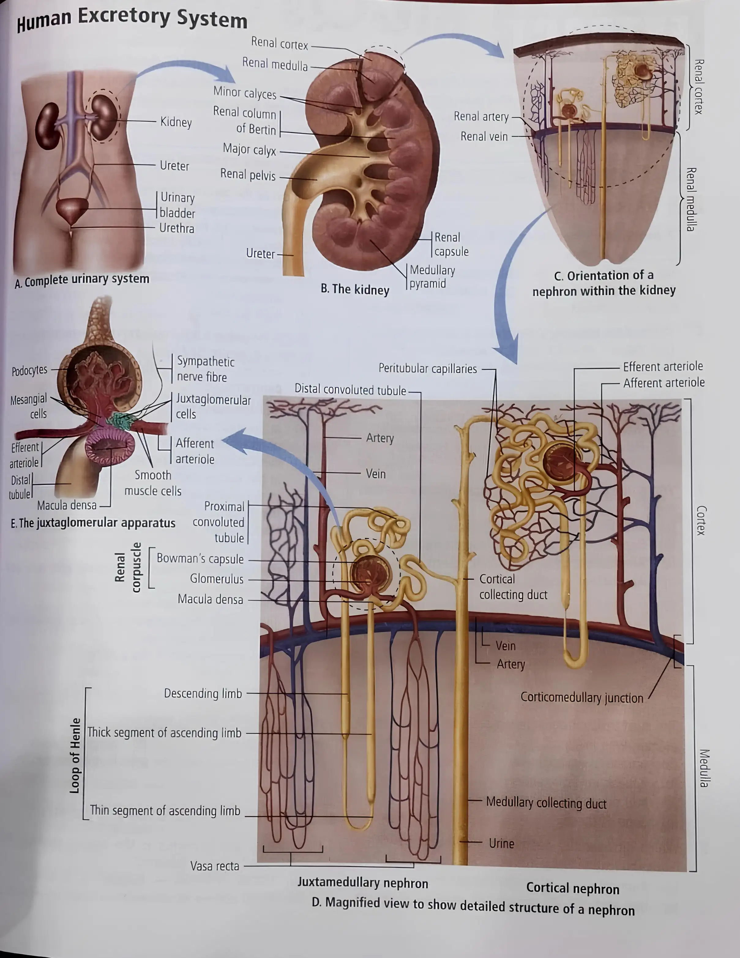
- Includes kidneys, ureters, bladder, and urethra.
- Kidney has outer renal cortex and inner renal medulla.
- Contains renal pelvis and calyces for urine collection.
- Nephrons are filtering units in cortex and medulla.
- Nephron parts: Bowman's capsule, glomerulus, tubules, collecting ducts.
- Blood enters nephron via arterioles, filtered in glomerulus.
- Filtrate passes tubules; useful substances reabsorbed.
- Waste and excess fluid form urine, collected in ducts to renal pelvis.
- Urine flows through ureters to bladder, exits via urethra.
Memory Aids
- Nephron = "Kidney's filtering unit"
- Flow: Blood → Glomerulus → Tubules → Urine
Quick Checkpoints
- What are the main parts of the excretory system?
- Where are nephrons located?
- What is the function of Bowman's capsule?
- How is urine formed and transported?
NEET Zoology — ALL CHAPTERS
1
Structural Organisation in Animals
2
Breathing and Exchange of Gases
3
Body Fluids and Circulation
4
Excretory Products and their Elimination
5
Locomotion and Movement
6
Neural Control and Coordination
7
Human Reproduction
8
Reproductive Health
9
Evolution
10
Microbes in Human Welfare
11
Organism and Population
12
Ecosystem
13
Biodiversity and Conservation