Detailed Structure and Anatomy of Dicot Leaves
Fundamental Characteristics of Dicotyledonous Plants
Overview of Dicotyledons and Their Basic Features
Dicotyledons, commonly called dicots, belong to the flowering plants category within the Plant Kingdom. They are distinguished by having two embryonic seed leaves, known as cotyledons. Presently, over 175,000 species of dicots have been identified worldwide. Typical examples include plants such as roses, magnolias, and geraniums.
These plants exhibit clear differentiation into roots, stems, and leaves. Their flowers are composed of petals, sepals, pistils, and stamens. The leaves possess a sophisticated vascular network that facilitates the transport of nutrients and food throughout the plant body.
Example: Identify whether a sunflower, which has two cotyledons in its seed, is a dicot or monocot and justify your answer.
Solution:
Sunflower seeds contain two cotyledons, which is a defining feature of dicots. Additionally, sunflower leaves have a reticulate venation pattern typical of dicots. Therefore, sunflower is classified as a dicotyledonous plant.
Structural Composition of a Dicot Leaf
Understanding the Dorsiventral Leaf Architecture
A dicot leaf is dorsiventral, meaning it has two distinct surfaces: the upper (adaxial) and the lower (abaxial) surfaces. These surfaces differ in both appearance and internal structure, adapting the leaf for efficient photosynthesis and gas exchange.
The leaf is primarily composed of three layers: the epidermis, the mesophyll, and the vascular system. The epidermis covers both surfaces and is coated with a thin cuticle that protects the leaf from physical damage and water loss.
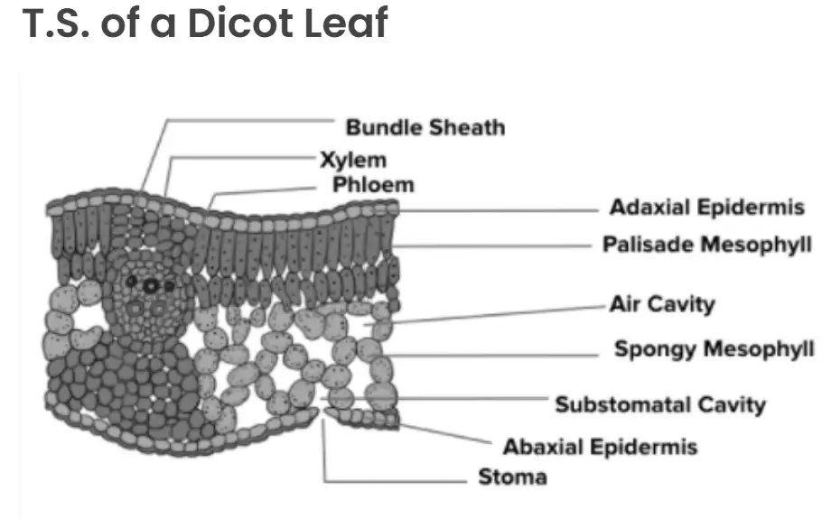
Diagram: Transverse section of a dicot leaf
Example: Explain why the cuticle layer on the epidermis is essential for dicot leaves.
Solution:
The cuticle reduces water loss by evaporation, helping the plant conserve moisture.
It acts as a barrier against mechanical injury and pathogen invasion.
Its thinness allows light to penetrate for photosynthesis while providing protection.
Detailed Layers Within the Dicot Leaf
Mesophyll and Vascular Tissue Arrangement
The mesophyll, located between the upper and lower epidermis, consists of parenchyma cells rich in chloroplasts, which are vital for photosynthesis. It is divided into two distinct types:
Palisade Parenchyma: Positioned just beneath the upper epidermis, this layer contains elongated, tightly packed cells arranged vertically to maximize light absorption.
Spongy Parenchyma: Found closer to the lower epidermis, this layer has loosely arranged, rounded cells with numerous air spaces facilitating gas exchange.
The lower surface of the leaf generally contains more stomata than the upper surface, aiding in efficient gas exchange. The vascular bundles, comprising xylem and phloem, are embedded within the leaf veins and midrib, surrounded by protective bundle sheath cells made of parenchyma layers.
Quick Reference: Key Features of Dicot Leaf Anatomy
Component | Description | Function |
|---|---|---|
Upper Epidermis | Single layer of cells with thin cuticle | Protection and light penetration |
Palisade Parenchyma | Elongated, tightly packed cells beneath upper epidermis | Primary site of photosynthesis |
Spongy Parenchyma | Loosely arranged cells with air spaces | Facilitates gas exchange |
Lower Epidermis | Contains more stomata than upper epidermis | Regulates gas exchange and transpiration |
Vascular Bundles | Composed of xylem and phloem, surrounded by bundle sheath | Transport of water, minerals, and food |
Glossary of Important Terms
Term | Definition |
|---|---|
Adaxial Surface | The upper side of a leaf facing the stem |
Abaxial Surface | The lower side of a leaf, opposite to the adaxial |
Cuticle | A waxy protective layer covering the epidermis |
Mesophyll | The inner tissue of a leaf where photosynthesis occurs |
Palisade Parenchyma | Layer of elongated cells rich in chloroplasts beneath upper epidermis |
Spongy Parenchyma | Layer of loosely arranged cells with air spaces for gas exchange |
Stomata | Small pores on leaf surfaces for gas exchange |
Vascular Bundle | Strands of xylem and phloem tissues for transport |
Bundle Sheath | Protective layer of cells surrounding vascular bundles |
Dicotyledon | Plant group with two seed leaves in the embryo |
Frequently Asked Questions
What distinguishes a dicot leaf from a monocot leaf?
Dicot leaves are dorsiventral with distinct upper and lower surfaces and have a reticulate venation pattern, whereas monocot leaves are usually isobilateral with parallel venation.
Why are there more stomata on the lower surface of a dicot leaf?
The lower surface is less exposed to direct sunlight, reducing water loss, so it contains more stomata to facilitate gas exchange efficiently without excessive transpiration.
What is the role of palisade parenchyma in a dicot leaf?
Palisade parenchyma cells are packed with chloroplasts and are the primary site for photosynthesis, capturing maximum light energy.
How do vascular bundles support leaf function?
Vascular bundles transport water, minerals, and food throughout the leaf, supporting metabolic activities and structural integrity.
What purpose do bundle sheath cells serve?
Bundle sheath cells protect vascular tissues and help regulate the movement of substances between the vascular bundles and surrounding cells.