Comprehensive Overview of the Spinal Cord
Fundamental Structure and Organization of the Spinal Cord
Basic Anatomy and Segmentation
The spinal cord is a crucial component of the central nervous system, extending as a cylindrical structure from the medulla oblongata through the vertebral canal. In adults, it typically measures around 38 to 42 centimeters in length and approximately 2 centimeters in diameter, serving as the primary conduit between the brain and the rest of the body.
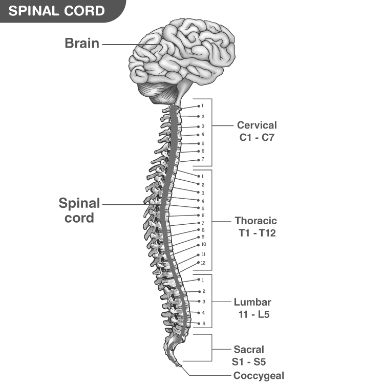
This structure is divided into five distinct regions: cervical, thoracic, lumbar, sacral, and coccygeal segments. Each segment gives rise to paired spinal nerves that exit the vertebral column, with 8 cervical, 12 thoracic, 5 lumbar, 5 sacral, and 1 coccygeal pairs in total. These nerves facilitate communication between the central nervous system and specific body areas.
The spinal cord's internal composition includes grey matter centrally located in a butterfly shape, surrounded by white matter. The grey matter contains neuron cell bodies and is organized into dorsal and ventral horns, while the white matter consists of myelinated axons forming ascending and descending tracts for signal transmission.
Illustration of the spinal cord within the vertebral column showing segmented nerves and surrounding bones.
Protective Layers and Internal Composition
The spinal cord is enveloped by three protective membranes known as meninges: dura mater, arachnoid mater, and pia mater. The dura mater is the tough outermost layer, consisting of periosteal and meningeal layers separated by the epidural space. Beneath it lies the arachnoid mater, and the innermost pia mater closely adheres to the spinal cord surface.
The subarachnoid space between the arachnoid and pia mater contains cerebrospinal fluid (CSF), which cushions the spinal cord and facilitates nutrient transport. The central canal within the grey matter also contains CSF, maintaining the internal environment of the spinal cord.
White matter tracts surrounding the grey matter are responsible for transmitting sensory and motor signals between the brain and peripheral nerves. Ascending tracts carry sensory information to the brain, while descending tracts convey motor commands to the body.
Example Problem
Explain the role of the subarachnoid space in spinal cord protection and identify which fluid it contains.
Solution:
The subarachnoid space lies between the arachnoid mater and pia mater.
It contains cerebrospinal fluid (CSF), which acts as a cushion to absorb shocks and protect the spinal cord from injury.
CSF also helps in nutrient transport and waste removal within the central nervous system.
Spinal Cord Nerves and Their Functional Roles
Classification and Distribution of Spinal Nerves
The spinal cord connects to the body through 31 pairs of spinal nerves, each attached via two roots: the dorsal (sensory) root and the ventral (motor) root. The dorsal roots transmit sensory information from the body to the spinal cord, while the ventral roots carry motor commands from the spinal cord to muscles.
These nerves are categorized based on the spinal segments they emerge from:
Cervical nerves (8 pairs): Originate from the neck region (C1-C8).
Thoracic nerves (12 pairs): Emerge from the chest area (T1-T12).
Lumbar nerves (5 pairs): Arise from the lower back (L1-L5).
Sacral nerves (5 pairs): Emanate from the sacrum (S1-S5), a fused bone at the base of the spine.
Coccygeal nerve (1 pair): Comes from the tailbone region.
Each spinal nerve innervates a specific skin region called a dermatome, enabling precise sensory and motor control.
Example Problem
A patient has lost sensation in the skin area supplied by the L3 spinal nerve. Identify the spinal region affected and the type of nerve root involved in carrying sensory signals.
Solution:
The L3 nerve belongs to the lumbar region of the spinal cord.
The dorsal root of the L3 spinal nerve carries sensory impulses from the skin to the spinal cord.
Therefore, damage to the dorsal root at L3 results in loss of sensation in its dermatome.
Motor and Sensory Pathways in Spinal Nerves
Spinal nerves serve as communication channels, transmitting motor commands from the central nervous system to muscles and sensory information from receptors back to the CNS. The ventral roots contain axons of motor neurons that control skeletal and smooth muscles, while the dorsal roots carry sensory fibers from receptors detecting stimuli such as touch, pain, and temperature.
This bidirectional flow enables reflex actions and voluntary movements, maintaining body posture and responding to environmental changes.
Example Problem
Describe the pathway of a reflex action initiated by touching a hot object, involving spinal nerves.
Solution:
Sensory receptors in the skin detect the heat stimulus.
Sensory impulses travel via the dorsal root of the spinal nerve to the spinal cord.
Interneurons in the grey matter process the information and activate motor neurons.
Motor impulses exit through the ventral root to the muscles, causing withdrawal of the hand.
Spinal Cord Injuries and Their Consequences
Types and Effects of Spinal Cord Damage
Injuries to the spinal cord or its nerves can cause irreversible damage, severely impairing motor and sensory functions below the injury site. The extent of disability depends on the injury's location and severity.
Complete injuries result in total loss of movement and sensation below the affected area, while incomplete injuries allow partial preservation of these functions. Such trauma can also affect reflexes, posture, and autonomic functions like breathing and bladder control.
Example Problem
A person suffers a spinal injury at the thoracic level resulting in paralysis of the lower limbs but intact arm movement. Classify the type of paralysis and explain why the arms are unaffected.
Solution:
The paralysis affecting only the lower limbs is called paraplegia.
The thoracic injury spares the cervical spinal cord segments that control arm movement.
Thus, motor and sensory functions of the arms remain intact.
Paralysis Types and Associated Symptoms
Two primary forms of paralysis arise from spinal cord injuries:
Tetraplegia (Quadriplegia): Involves partial or complete loss of function in all four limbs and torso, typically due to cervical spinal cord damage.
Paraplegia: Affects only the lower limbs and lower body, sparing the arms, usually resulting from injuries below the cervical region.
Additional symptoms may include altered reflexes, loss of sensation, muscle spasms, respiratory difficulties, and changes in sexual function.
Example Problem
Identify the type of paralysis and possible symptoms if a patient loses voluntary control of all limbs and experiences breathing difficulties.
Solution:
The condition is tetraplegia, indicating cervical spinal cord involvement.
Breathing problems arise due to impaired control of respiratory muscles.
Other symptoms may include loss of sensation and muscle spasms.
Essential Functions and Physiological Roles of the Spinal Cord
Communication and Reflex Coordination
The spinal cord acts as a vital communication highway, linking the brain with the peripheral nervous system. It transmits sensory data from receptors to the brain and conveys motor commands from the brain to muscles, enabling coordinated movement and posture maintenance.
Additionally, the spinal cord orchestrates reflexes—automatic responses to stimuli—through local neural circuits, allowing rapid reactions without brain involvement.
Example Problem
Explain how the spinal cord facilitates a knee-jerk reflex when the patellar tendon is tapped.
Solution:
The tap stretches receptors in the patellar tendon.
Sensory neurons send impulses via dorsal roots to the spinal cord.
Motor neurons in the ventral horn are activated, causing the quadriceps muscle to contract.
This results in the leg kicking forward, completing the reflex arc.
Structural Support and Electrical Insulation
The spinal cord provides structural integrity to the body, supporting posture and enabling flexible movements. The white matter contains myelin, a fatty substance that electrically insulates axons, enhancing the speed and efficiency of nerve signal transmission.
This insulation is critical for rapid communication between the brain and peripheral nerves, ensuring timely responses to sensory inputs and motor commands.
Quick Reference: Spinal Cord Overview
Aspect | Details |
|---|---|
Length | Approximately 40 cm in adults |
Width | About 2 cm |
Regions | Cervical, Thoracic, Lumbar, Sacral, Coccygeal |
Number of Spinal Nerves | 31 pairs (8 cervical, 12 thoracic, 5 lumbar, 5 sacral, 1 coccygeal) |
Protective Layers | Dura mater, Arachnoid mater, Pia mater |
Grey Matter | Contains neuron cell bodies; shaped like a butterfly |
White Matter | Myelinated axons forming ascending and descending tracts |
Functions | Signal transmission, reflex coordination, structural support |
Types of Paralysis | Tetraplegia and Paraplegia |
Fluid in Central Canal | Cerebrospinal Fluid (CSF) |
Glossary of Key Terms
Term | Definition |
|---|---|
Spinal Cord | Long, cylindrical structure connecting brain to body, part of CNS |
Medulla Oblongata | Lower part of the brainstem from which the spinal cord arises |
Vertebral Column | Series of vertebrae protecting the spinal cord |
Grey Matter | Region containing neuron cell bodies within the spinal cord |
White Matter | Myelinated axons transmitting signals in the spinal cord |
Dorsal Root | Spinal nerve root carrying sensory information to the CNS |
Ventral Root | Spinal nerve root carrying motor commands from the CNS |
Dermatome | Skin area innervated by a specific spinal nerve |
Meninges | Protective membranes surrounding the spinal cord |
Cerebrospinal Fluid (CSF) | Fluid cushioning the brain and spinal cord |
Frequently Asked Questions
What is the primary role of the spinal cord?
The spinal cord serves as the main communication pathway between the brain and the rest of the body, transmitting sensory and motor signals.
How many pairs of spinal nerves are there, and how are they distributed?
There are 31 pairs of spinal nerves: 8 cervical, 12 thoracic, 5 lumbar, 5 sacral, and 1 coccygeal pair.
What are the main protective layers of the spinal cord?
The spinal cord is protected by three meninges: dura mater, arachnoid mater, and pia mater, with cerebrospinal fluid cushioning between arachnoid and pia mater.
What is the difference between tetraplegia and paraplegia?
Tetraplegia affects all four limbs and torso, usually from cervical injuries, while paraplegia affects only the lower limbs, sparing the arms.
How does the spinal cord contribute to reflex actions?
The spinal cord processes reflexes locally by receiving sensory input and sending immediate motor output without involving the brain, enabling quick responses.