Comprehensive Overview of Female Gamete Formation
Understanding the Formation of Female Gametes
Fundamentals of Female Gamete Development
Female gamete formation, scientifically termed oogenesis, is the biological process through which ova or egg cells develop. These ova represent the female reproductive cells, each containing a single set of chromosomes, making them haploid. The term "egg" can refer to various developmental stages depending on the organism. For instance, in birds, the egg encompasses the entire developmental journey until hatching, whereas in placental mammals, the egg is fertilized and begins cell division, transitioning beyond the egg stage.
Diagram depicting the stages of oogenesis
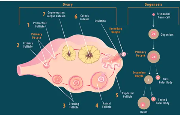
Oogenesis is distinct from spermatogenesis, the male counterpart of gamete formation. While spermatogenesis produces sperm continuously after puberty, oogenesis initiates during fetal development and proceeds through specific stages before and after birth.
Example Problem
Question: Explain why the ovum must be haploid and how this is ensured during oogenesis.
Solution:
The ovum must be haploid to ensure that upon fertilization, the resulting zygote has the correct diploid number of chromosomes.
During oogenesis, meiosis reduces the chromosome number from diploid (2n) to haploid (n).
Specifically, meiosis I separates homologous chromosomes, and meiosis II separates sister chromatids, resulting in a haploid ovum.
This reduction prevents chromosome doubling in each generation, maintaining genetic stability.
Stages and Progression of Oocyte Development
Initial Phase: Prenatal Development of Oocytes
Oogenesis begins in the female fetus before birth. The germ cells, called oogonia, multiply by mitosis and then differentiate into primary oocytes. These primary oocytes enter the first meiotic division but halt at prophase I, remaining arrested until puberty. Surrounding these oocytes, follicular cells multiply to form granulosa cells, which secrete glycoproteins creating a protective layer called the zona pellucida.
Example Problem
Question: Describe the significance of the arrest of primary oocytes during the prenatal stage.
Solution:
The arrest at prophase I preserves the oocytes in a dormant state until hormonal signals trigger further development.
This pause allows the female to be born with a finite number of oocytes, which will mature over her reproductive lifespan.
It prevents premature completion of meiosis, ensuring oocytes are available for ovulation during reproductive years.
Secondary Phase: Formation of Antral Follicles
As the female matures, some primary oocytes resume development monthly. Fluid accumulates between granulosa cells, merging into a cavity called the antrum, forming secondary follicles. These follicles grow under the influence of hormones such as follicle-stimulating hormone (FSH) and luteinizing hormone (LH), preparing the oocyte for ovulation.
Example Problem
Question: How do hormones regulate the growth of secondary follicles during the antral stage?
Solution:
FSH stimulates granulosa cells to proliferate and produce estrogen, promoting follicle growth.
LH supports theca cells to produce androgens, which granulosa cells convert to estrogen.
The rising estrogen levels prepare the follicle and oocyte for the next stage of development.
Final Phase: Pre-Ovulatory Maturation
Triggered by a surge in LH, the primary oocyte completes meiosis I, producing two haploid cells of unequal size: a large secondary oocyte and a small polar body. The polar body typically degenerates and does not contribute to fertilization. The secondary oocyte begins meiosis II but halts at metaphase II, awaiting fertilization to complete the process.
Example Problem
Question: Explain the role of the polar body formed during the pre-ovulatory stage.
Solution:
The polar body contains a haploid set of chromosomes but minimal cytoplasm.
Its formation ensures that the secondary oocyte retains most of the cytoplasm, essential for early embryonic development.
Polar bodies usually degenerate and do not participate in fertilization.
Ovulation and Fertilization Processes
Release of the Secondary Oocyte: Ovulation
During each menstrual cycle, typically one follicle matures fully while others regress. The mature follicle releases the secondary oocyte into the fallopian tube in a process called ovulation. At this point, the secondary oocyte is arrested in metaphase II and will only complete meiosis II if fertilization occurs.
Example Problem
Question: What happens to the secondary oocyte if fertilization does not take place after ovulation?
Solution:
If fertilization does not occur within approximately 24 hours, the secondary oocyte degenerates.
It remains arrested in metaphase II and does not complete meiosis.
This degeneration prevents the formation of an embryo without fertilization.
Completion of Meiosis and Fertilization
Fertilization triggers the secondary oocyte to complete meiosis II, producing a mature ovum and a second polar body. The fusion of the haploid sperm and ovum nuclei restores the diploid chromosome number, initiating embryonic development.
Example Problem
Question: How does fertilization influence the completion of meiosis in the secondary oocyte?
Solution:
The sperm entry activates the secondary oocyte to complete meiosis II.
This results in the formation of the mature ovum and a second polar body.
The mature ovum then fuses with the sperm nucleus to form a diploid zygote.
Quick Reference: Key Points on Female Gamete Formation
Aspect | Description |
|---|---|
Starting Cell | Oogonium (diploid germ cell) |
Location | Outer layers of the ovary |
Stages | Pre-natal, Antral, Pre-ovulatory |
Meiosis Arrest Points | Prophase I (primary oocyte), Metaphase II (secondary oocyte) |
Hormonal Influence | FSH and LH regulate follicle growth and ovulation |
Polar Bodies | Small haploid cells that do not develop into ova |
Ovulation | Release of secondary oocyte into fallopian tube |
Fertilization Effect | Completion of meiosis II and formation of zygote |
Difference from Spermatogenesis | Oogenesis begins before birth; spermatogenesis starts at puberty |
Oocyte Quantity | Finite number established before birth |
Glossary of Terms Related to Female Gamete Formation
Term | Definition |
|---|---|
Oogenesis | Process of female gamete (ovum) formation |
Oogonium | Diploid germ cell that divides to form primary oocytes |
Primary Oocyte | Cell arrested in prophase I of meiosis, precursor to ovum |
Secondary Oocyte | Haploid cell formed after meiosis I, arrested in metaphase II |
Polar Body | Small haploid cell produced during meiosis, usually degenerates |
Granulosa Cells | Follicular cells surrounding the oocyte, secrete zona pellucida |
Zona Pellucida | Glycoprotein layer surrounding the oocyte |
Antrum | Fluid-filled cavity in secondary follicles |
Follicle-Stimulating Hormone (FSH) | Hormone stimulating follicle growth |
Luteinizing Hormone (LH) | Hormone triggering ovulation and meiosis completion |
Frequently Asked Questions on Female Gamete Formation
What is the primary purpose of oogenesis?
Oogenesis produces haploid female gametes (ova) necessary for sexual reproduction, ensuring genetic diversity and chromosome number stability.
At which stage does the primary oocyte pause during development?
The primary oocyte halts at prophase I of meiosis during fetal development and remains arrested until puberty.
How do hormones influence the oogenesis process?
FSH promotes follicle growth and estrogen production, while LH triggers ovulation and completion of meiosis I in the oocyte.
What happens to the secondary oocyte if fertilization does not occur?
It degenerates approximately 24 hours after ovulation, remaining arrested in metaphase II without completing meiosis.
How does oogenesis differ from spermatogenesis?
Oogenesis begins before birth with a finite number of oocytes, while spermatogenesis starts at puberty and continues throughout life producing sperm continuously.