Comparative Anatomy of Monocot and Dicot Plants
Root Structure Differences in Monocot and Dicot Plants
Characteristics of Dicot Roots
Dicotyledonous plants develop a taproot system, which is a prominent central root with smaller lateral branches. The outermost layer of the root is the epidermis, which often extends into root hairs to increase surface area for absorption. Beneath the epidermis lies the cortex, composed of loosely packed parenchyma cells with intercellular spaces facilitating gas exchange.
The innermost layer of the cortex is the endodermis, consisting of tightly packed, barrel-shaped cells that regulate water movement. Inside the endodermis is the pericycle, a few layers of thick-walled parenchyma cells that give rise to lateral roots. The vascular tissue in dicot roots includes two to four xylem and phloem bundles arranged alternately, separated by parenchymatous conjunctive tissue. Unlike monocots, the central pith is not well-defined in dicot roots. During secondary growth, the cambium forms between xylem and phloem, contributing to the thickening of the root. The pericycle, vascular bundles, and pith collectively form the stele in dicots.
Example: A dicot root has 3 xylem bundles and 3 phloem bundles arranged alternately. If the diameter of the root is 2 cm, estimate the approximate spacing between two adjacent xylem bundles assuming they are evenly spaced around the center.
Solution: The xylem bundles are arranged in a circle inside the root. The circumference of the circle where xylem bundles lie can be approximated by the root's internal vascular cylinder circumference. Assuming the vascular cylinder diameter is about 1.5 cm, the circumference is:
\[ C = \pi \times d = \pi \times 1.5 = 4.71 \text{ cm} \]
Since there are 3 xylem bundles evenly spaced, the distance between adjacent bundles along the circumference is:
\[ \frac{4.71}{3} = 1.57 \text{ cm} \]
Thus, the xylem bundles are approximately 1.57 cm apart along the vascular ring.
Features of Monocot Roots
Monocotyledonous plants possess an adventitious root system, which arises from stem tissues rather than a primary root. The root anatomy resembles that of dicots in having an epidermis, cortex, endodermis, pericycle, vascular bundles, and pith, but with notable distinctions. The pith in monocot roots is large and conspicuous, unlike the indistinct pith in dicots. The number of xylem bundles is generally six or more, arranged in a ring around the pith. Secondary growth is absent in monocot roots, so they do not thicken over time as dicot roots do.
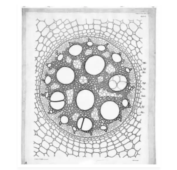
Cross section of a monocot root
Example: A monocot root has 8 xylem bundles arranged around a large pith. If the root diameter is 3 cm and the pith diameter is 1 cm, calculate the approximate distance between two adjacent xylem bundles along the vascular ring.
Solution: The vascular ring diameter is approximately the root diameter minus the pith diameter:
\[ d = 3 - 1 = 2 \text{ cm} \]
The circumference of the vascular ring is:
\[ C = \pi \times d = \pi \times 2 = 6.28 \text{ cm} \]
With 8 xylem bundles evenly spaced, the distance between adjacent bundles is:
\[ \frac{6.28}{8} = 0.785 \text{ cm} \]
Therefore, the xylem bundles are about 0.785 cm apart along the ring.
Stem Anatomy: Contrasting Monocot and Dicot Structures
Structural Traits of Dicot Stems
Dicot stems are typically solid and exhibit a well-organized internal structure. The outermost layer is the epidermis, which is coated with a thin cuticle to prevent water loss. This layer contains trichomes and stomata for protection and gas exchange. Beneath the epidermis lies the cortex, which is divided into three sublayers: the hypodermis made of collenchyma cells providing mechanical support, the cortical parenchyma cells, and the innermost endodermis.
Next is the pericycle, composed of semi-lunar sclerenchyma patches that add strength. The vascular bundles in dicot stems are arranged in a distinct ring, each bundle being conjoint (containing both xylem and phloem), open (with cambium present), and having endarch protoxylem (protoxylem towards the outside). The central region, called the pith, consists of parenchyma cells and is clearly visible.
Example: In a young dicot stem, the vascular bundles are arranged in a ring with 12 bundles. If the stem diameter is 4 cm, estimate the average distance between two adjacent vascular bundles along the ring.
Solution: The vascular bundles lie in a ring inside the stem. Assuming the ring diameter is about 3.5 cm (slightly less than stem diameter), the circumference is:
\[ C = \pi \times 3.5 = 10.995 \text{ cm} \]
Dividing by 12 bundles:
\[ \frac{10.995}{12} = 0.916 \text{ cm} \]
So, the bundles are approximately 0.916 cm apart along the ring.
Distinctive Features of Monocot Stems
Monocot stems are generally hollow and lack secondary growth, which means they do not thicken over time. The cortex hypodermis is composed of sclerenchymatous cells, providing rigidity. Unlike dicots, monocot vascular bundles are numerous, scattered throughout the ground tissue, and are conjoint but closed (no cambium present). Additionally, monocot stems lack phloem parenchyma cells, which are present in dicots.
Example: A monocot stem has 30 scattered vascular bundles within a stem diameter of 5 cm. If the bundles are uniformly distributed, estimate the average area available per vascular bundle.
Solution: The cross-sectional area of the stem is:
\[ A = \pi r^2 = \pi \times \left(\frac{5}{2}\right)^2 = \pi \times 2.5^2 = 19.635 \text{ cm}^2 \]
Dividing by 30 bundles:
\[ \frac{19.635}{30} = 0.6545 \text{ cm}^2 \]
Each vascular bundle occupies approximately 0.6545 cm² of area.
Leaf Anatomy: Comparing Monocot and Dicot Leaves
Structure of Dicot Leaves
Dicot leaves exhibit reticulate venation, where veins form a network. The leaf lamina consists of three main layers: the epidermis, mesophyll, and vascular system. The epidermis is covered by a protective cuticle and contains stomata for gas exchange. The lower (abaxial) epidermis usually has more stomata than the upper (adaxial) epidermis, which sometimes lacks stomata entirely.
The mesophyll is differentiated into palisade parenchyma, which is tightly packed and rich in chloroplasts for photosynthesis, and spongy parenchyma, which has loosely arranged cells with air spaces to facilitate gas diffusion. Vascular bundles, surrounded by bundle sheath cells, form the veins and midrib of the leaf.
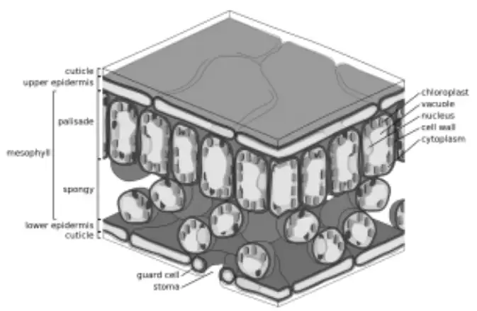
Anatomy of a dicot leaf
Example: A dicot leaf has 60% palisade parenchyma and 40% spongy parenchyma in its mesophyll. If the total mesophyll thickness is 0.5 mm, calculate the thickness of each parenchyma layer.
Solution: Thickness of palisade parenchyma:
\[ 0.6 \times 0.5 = 0.3 \text{ mm} \]
Thickness of spongy parenchyma:
\[ 0.4 \times 0.5 = 0.2 \text{ mm} \]
Thus, palisade layer is 0.3 mm thick and spongy layer is 0.2 mm thick.
Monocot Leaf Characteristics
Monocot leaves are distinguished by parallel venation, where veins run side by side along the length of the leaf. Both the upper (adaxial) and lower (abaxial) epidermis contain stomata, unlike dicots where the upper epidermis may lack them. The mesophyll is not differentiated into palisade and spongy layers; instead, it consists of uniform parenchyma cells.
Unique to monocots are bulliform cells, large, empty cells derived from the adaxial epidermis. These cells help the leaf curl during dry conditions to reduce water loss by minimizing exposed surface area.
Example: A monocot leaf has bulliform cells that occupy 15% of the adaxial epidermis area. If the total adaxial epidermis area is 2 cm², find the area covered by bulliform cells.
Solution: Area of bulliform cells:
\[ 0.15 \times 2 = 0.3 \text{ cm}^2 \]
Therefore, bulliform cells cover 0.3 cm² of the adaxial epidermis.
Summary Table: Key Differences Between Monocots and Dicots
Feature | Dicot Plants | Monocot Plants |
|---|---|---|
Root System | Taproot system with indistinct pith | Adventitious roots with large, distinct pith |
Number of Xylem Bundles in Root | 2 to 4 | 6 or more |
Secondary Growth | Present (cambium active) | Absent |
Stem Structure | Solid stem with vascular bundles in a ring | Hollow stem with scattered vascular bundles |
Vascular Bundle Type | Conjoint, open, with cambium | Conjoint, closed, no cambium |
Leaf Venation | Reticulate (net-like) | Parallel |
Mesophyll | Differentiated into palisade and spongy parenchyma | Undifferentiated parenchyma |
Stomata Distribution | More on abaxial surface; sometimes absent on adaxial | Present on both adaxial and abaxial surfaces |
Bulliform Cells | Absent | Present, aid in leaf folding |
Phloem Parenchyma | Present | Absent |
Glossary of Important Terms
Term | Definition |
|---|---|
Adventitious Roots | Roots that develop from parts other than the primary root, such as stems or leaves. |
Cambium | A layer of meristematic tissue responsible for secondary growth in plants. |
Endodermis | The innermost layer of the cortex that regulates water and mineral movement into the vascular tissue. |
Mesophyll | The photosynthetic tissue in leaves, consisting of palisade and spongy parenchyma cells. |
Parenchyma | Fundamental plant tissue composed of living cells that perform various functions including storage and photosynthesis. |
Pericycle | A layer of cells just inside the endodermis that can give rise to lateral roots. |
Pith | The central part of the stem or root, composed mainly of parenchyma cells. |
Reticulate Venation | A leaf vein pattern where veins form a network, typical of dicots. |
Secondary Growth | Increase in thickness or girth of plant organs due to cambium activity. |
Vascular Bundle | Strands of xylem and phloem tissues that transport water, nutrients, and food in plants. |
Frequently Asked Questions
What is the main difference between monocot and dicot root systems?
Dicots have a taproot system with a small or indistinct pith, while monocots have adventitious roots with a large, distinct pith.
Why do monocot stems lack secondary growth?
Monocot stems do not have a vascular cambium, which is essential for secondary growth, so they remain the same thickness throughout their life.
How does leaf venation differ between monocots and dicots?
Monocot leaves have parallel venation with veins running side by side, whereas dicot leaves show reticulate venation forming a network.
What role do bulliform cells play in monocot leaves?
Bulliform cells help monocot leaves curl during dry conditions to reduce water loss by minimizing exposed surface area.
Can dicot stems have scattered vascular bundles like monocots?
No, dicot stems have vascular bundles arranged in a ring, which is a key distinguishing feature from monocots.