Comprehensive Guide to Breathing and Gas Exchange
Overview of the Human Respiratory Framework
Structure and Function of Respiratory Organs
Energy production in the body depends on breaking down nutrients such as carbohydrates, fats, and proteins. This process requires oxygen and produces carbon dioxide as a waste product. To maintain life, organisms must continuously exchange these gases with their environment, a process known as respiration.
Different animals have evolved specialized organs for this gas exchange. For example, earthworms use their moist skin, amphibians like frogs rely on their skin and lungs, while humans possess a complex respiratory system designed for efficient gas transfer.
In humans, the respiratory tract begins at the nostrils and continues through the pharynx, larynx, trachea, bronchi, bronchioles, and finally the alveoli, where gas exchange occurs. The lungs, protected by the pleural membranes and cushioned by pleural fluid, reside within the thoracic cavity formed by the ribs, sternum, vertebral column, and diaphragm.
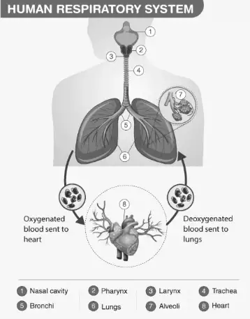
Diagram illustrating the human respiratory system
Example Problem
Question: Identify the sequence of air passage from the nostrils to the alveoli in the human respiratory system.
Answer:
Air enters through the nostrils.
Passes into the pharynx, a shared pathway for food and air.
Moves through the larynx, where the epiglottis prevents food from entering the airway.
Travels down the trachea, which bifurcates at the fifth thoracic vertebra.
Enters the primary bronchi (right and left), then into secondary and tertiary bronchi.
Reaches the bronchioles, which terminate in alveolar sacs.
Mechanics of Breathing and Lung Volumes
How Air Moves In and Out of the Lungs
Breathing involves two main phases: inspiration and expiration. During inspiration, the diaphragm and external intercostal muscles contract, expanding the thoracic cavity and lowering the pressure inside the lungs compared to the atmosphere, causing air to flow in.
Expiration occurs when these muscles relax, reducing lung volume and increasing pressure, pushing air out. Additional muscles in the abdomen can assist in deep breathing. The normal respiratory rate ranges from 12 to 16 breaths per minute.
A spirometer is a device used to measure the volume of air inhaled and exhaled, helping assess lung function.
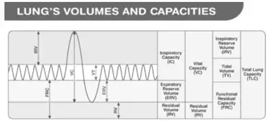
Illustration of various respiratory volumes and capacities
Example Problem
Question: Calculate the vital capacity of a person with tidal volume 450 ml, inspiratory reserve volume 2800 ml, and expiratory reserve volume 1100 ml.
Solution:
Vital capacity (VC) is the total volume of air that can be exhaled after a maximal inhalation, given by:
\[ VC = TV + IRV + ERV \]
Substituting the values:
\[ VC = 450 + 2800 + 1100 = 4350 \text{ ml} \]
Therefore, the vital capacity is 4350 ml or 4.35 liters.
Gas Exchange and Transport in the Body
Mechanisms of Oxygen and Carbon Dioxide Movement
Gas exchange occurs due to differences in partial pressures of oxygen and carbon dioxide between alveoli, blood, and body tissues. Oxygen diffuses from alveolar air into the blood, while carbon dioxide moves from blood to alveoli to be exhaled.
Oxygen is primarily transported bound to hemoglobin in red blood cells, forming oxyhemoglobin. Approximately 97% of oxygen is carried this way, with the remainder dissolved in plasma. The oxygen dissociation curve is sigmoidal, reflecting hemoglobin's affinity changes with oxygen concentration.
Carbon dioxide is transported in three forms: bound to hemoglobin as carbaminohemoglobin (20-25%), dissolved in plasma (7%), and mostly as bicarbonate ions (about 70%). The enzyme carbonic anhydrase catalyzes the reversible reaction:
\[ \mathrm{CO_2} + \mathrm{H_2O} \leftrightarrow \mathrm{H_2CO_3} \leftrightarrow \mathrm{HCO_3^-} + \mathrm{H^+} \]
This reaction facilitates efficient CO2 transport and release in the lungs.
Example Problem
Question: Explain why hemoglobin releases oxygen in tissues but binds oxygen in the lungs.
Answer:
In the lungs, high partial pressure of oxygen (\(pO_2\)) and low carbon dioxide levels favor oxygen binding to hemoglobin.
In tissues, lower \(pO_2\) and higher \(pCO_2\) cause hemoglobin to release oxygen for cellular use.
The presence of hydrogen ions and carbon dioxide shifts the oxygen dissociation curve to facilitate oxygen unloading.
Control and Disorders of the Respiratory System
Regulation of Breathing and Common Respiratory Conditions
Breathing is regulated by the respiratory centers located in the medulla oblongata and pons of the brainstem. These centers adjust the rate and depth of respiration based on the body's oxygen demand and carbon dioxide levels.
Several disorders can affect respiratory health:
Asthma: Characterized by inflammation and narrowing of airways, causing wheezing and breathing difficulty.
Emphysema: Damage to alveolar walls reduces surface area for gas exchange, often caused by smoking, leading to breathlessness.
Occupational Lung Diseases: Long-term exposure to dust and pollutants can cause lung inflammation, fibrosis, and impaired function.
Example Problem
Question: Describe the role of the medulla oblongata in respiratory regulation.
Answer:
The medulla oblongata contains the respiratory rhythm center that generates the basic breathing pattern.
It sends signals to respiratory muscles to control inhalation and exhalation.
The center adjusts breathing rate in response to changes in blood CO2 and O2 levels.
Quick Reference: Key Respiratory Volumes and Capacities
Term | Definition | Approximate Volume (ml) |
|---|---|---|
Tidal Volume (TV) | Air inhaled or exhaled during normal breathing | 500 |
Inspiratory Reserve Volume (IRV) | Additional air inhaled with force after normal inspiration | 2500–3000 |
Expiratory Reserve Volume (ERV) | Additional air exhaled with force after normal expiration | 1000–1100 |
Residual Volume (RV) | Air remaining in lungs after forced expiration | 1100–1200 |
Vital Capacity (VC) | Maximum air exhaled after maximum inhalation (TV + IRV + ERV) | 3500–4500 |
Total Lung Capacity (TLC) | Sum of vital capacity and residual volume | 4500–5700 |
Glossary of Essential Terms
Term | Meaning |
|---|---|
Alveoli | Microscopic air sacs in lungs where gas exchange occurs |
Bronchi | Large air passages that branch from the trachea into the lungs |
Diaphragm | Muscle that separates thoracic and abdominal cavities and aids breathing |
Epiglottis | Flap that prevents food from entering the windpipe during swallowing |
Hemoglobin | Protein in red blood cells that carries oxygen |
Inspiratory Reserve Volume | Extra air inhaled beyond normal breath |
Oxyhemoglobin | Hemoglobin bound to oxygen |
Residual Volume | Air left in lungs after forceful exhalation |
Respiratory Rate | Number of breaths per minute |
Vital Capacity | Maximum volume of air exhaled after a deep breath |
Frequently Asked Questions
What is the primary function of alveoli in the lungs?
Alveoli facilitate the exchange of oxygen and carbon dioxide between the air and blood due to their thin walls and large surface area.
How does the diaphragm contribute to breathing?
The diaphragm contracts to increase thoracic volume during inhalation and relaxes to decrease it during exhalation, driving air movement.
Why is hemoglobin important for oxygen transport?
Hemoglobin binds oxygen efficiently in the lungs and releases it in tissues where oxygen is needed, enhancing oxygen delivery.
What causes asthma and how does it affect breathing?
Asthma is caused by inflammation and narrowing of airways, leading to difficulty in breathing and wheezing.
How is carbon dioxide transported in the blood?
Carbon dioxide is transported as bicarbonate ions, bound to hemoglobin, and dissolved in plasma, with bicarbonate being the major form.