Comprehensive Overview of Human Eye Anatomy and Function
Fundamental Components and Roles of the Human Eye
External Features and Their Purposes
The human eye's outer visible parts serve crucial roles in protection, light regulation, and vision clarity. The sclera, a tough white layer, safeguards the delicate inner structures. Covering the sclera is the conjunctiva, a moist membrane that lubricates the eye and shields it from irritants by producing mucus and tears. The transparent cornea at the front acts as the primary refractive surface, bending incoming light rays. The iris, the colored ring, adjusts the pupil size to control light entry, while the pupil itself is the central opening allowing light to reach the retina.
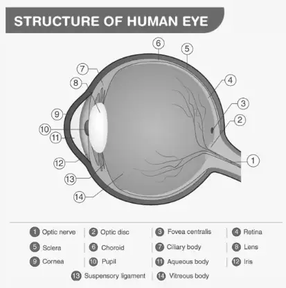
This structure helps the eye gather light, focus it, and send clear images to the brain.
Detailed structure of the human eye showing external parts
Example Problem
Calculate the diameter of the pupil if the iris adjusts it to allow 0.5 cm² of light to enter. Assume the pupil is circular.
Solution:
The area \( A \) of the pupil is given as \( 0.5 \text{ cm}^2 \). For a circle, \( A = \pi r^2 \).
Rearranging for radius \( r \):
\[ r = \sqrt{\frac{A}{\pi}} = \sqrt{\frac{0.5}{3.1416}} \approx \sqrt{0.159} \approx 0.399 \text{ cm} \]
Diameter \( d = 2r = 2 \times 0.399 = 0.798 \text{ cm} \).
Therefore, the pupil diameter is approximately \( 0.8 \text{ cm} \).
Internal Structures and Their Functions
Inside the eye, several specialized components work together to process visual information. The lens, a transparent biconvex structure, fine-tunes light refraction to focus images precisely on the retina. The retina, the innermost layer, contains photoreceptor cells that convert light into electrical signals. These signals travel via the optic nerve to the brain for interpretation. The aqueous humour, a clear fluid between the cornea and lens, nourishes the eye and maintains pressure, while the vitreous humour, a gel-like substance filling the space between the lens and retina, preserves the eye's shape and cushions internal parts.
Example Problem
If the focal length of the eye's lens is \( 2.5 \text{ cm} \), calculate the power of the lens in diopters.
Solution:
Power \( P \) in diopters is given by:
\[ P = \frac{100}{f} \]
where \( f \) is the focal length in centimeters.
Substituting \( f = 2.5 \text{ cm} \):
\[ P = \frac{100}{2.5} = 40 \text{ diopters} \]
The lens power is \( 40 \text{ diopters} \).
Unique Structural Characteristics of the Eye
Unlike a perfect sphere, the human eye consists of two fused segments forming an approximately spherical shape. This design allows for complex movements and precise focusing. The eye's muscles enable rotation within the eye socket, while the layered tissues and fluids maintain its shape and function. Understanding this composite structure is essential for grasping how vision is achieved and maintained.
Cross-sectional view showing the fused segments of the human eye
Example Problem
Explain why the human eye is not a perfect sphere and how this affects its function.
Answer:
The eye is composed of two fused segments rather than a perfect sphere, allowing for flexibility and movement.
This structure supports the attachment of muscles that control eye rotation and focus adjustment.
The shape aids in maintaining the correct focal length and image formation on the retina.
It also helps accommodate changes in pressure and volume within the eye.
Summary Table: Key Eye Structures and Their Roles
Eye Component | Description | Primary Function |
|---|---|---|
Sclera | White, tough outer layer | Protects inner eye parts |
Conjunctiva | Moist membrane covering sclera | Lubricates and protects eye surface |
Cornea | Transparent front layer | Refracts light entering the eye |
Iris | Colored ring around pupil | Controls pupil size to regulate light |
Pupil | Central aperture in iris | Allows light to reach retina |
Lens | Transparent biconvex structure | Focuses light on retina |
Retina | Light-sensitive inner layer | Converts light to nerve signals |
Optic Nerve | Nerve bundle at eye's back | Transmits visual information to brain |
Aqueous Humour | Clear fluid between cornea and lens | Nourishes eye and maintains pressure |
Vitreous Humour | Gel-like substance filling eye interior | Maintains eye shape and cushions parts |
Glossary of Essential Eye Anatomy Terms
Term | Definition |
|---|---|
Sclera | The white, fibrous outer layer of the eyeball providing protection. |
Conjunctiva | A thin, transparent membrane that covers the sclera and lubricates the eye. |
Cornea | The clear, curved front surface of the eye that refracts light. |
Iris | The colored part of the eye that controls the size of the pupil. |
Pupil | The opening in the center of the iris that allows light to enter. |
Lens | A transparent, flexible structure that focuses light onto the retina. |
Retina | The light-sensitive layer at the back of the eye that converts light into nerve signals. |
Optic Nerve | The nerve that transmits visual information from the retina to the brain. |
Aqueous Humour | The clear fluid between the cornea and lens that nourishes the eye. |
Vitreous Humour | The gel-like substance filling the eye's interior, maintaining its shape. |
Frequently Asked Questions on Eye Anatomy
What are the main external parts of the human eye?
The primary external components include the sclera, conjunctiva, cornea, iris, and pupil, each playing a role in protection, lubrication, light refraction, and regulation.
How does the conjunctiva contribute to eye health?
It keeps the eye surface moist, provides lubrication through mucus and tears, and acts as a barrier against dust and microbes.
What is the function of the iris and how is it structured?
The iris controls the pupil size to regulate light entry. It consists of two layers: the pigmented stroma and the pigmented epithelial cells.
Which internal parts of the eye are responsible for focusing light?
The cornea and the lens work together to refract and focus light precisely onto the retina for clear vision.
Why is the human eye not a perfect sphere?
Because it is formed by two fused segments, this shape allows for muscle attachment and eye movement, as well as maintaining proper focus and internal pressure.