Comprehensive Overview of the Human Ear: Structure and Functions
Detailed Anatomy of the Human Ear
External Ear Components and Their Roles
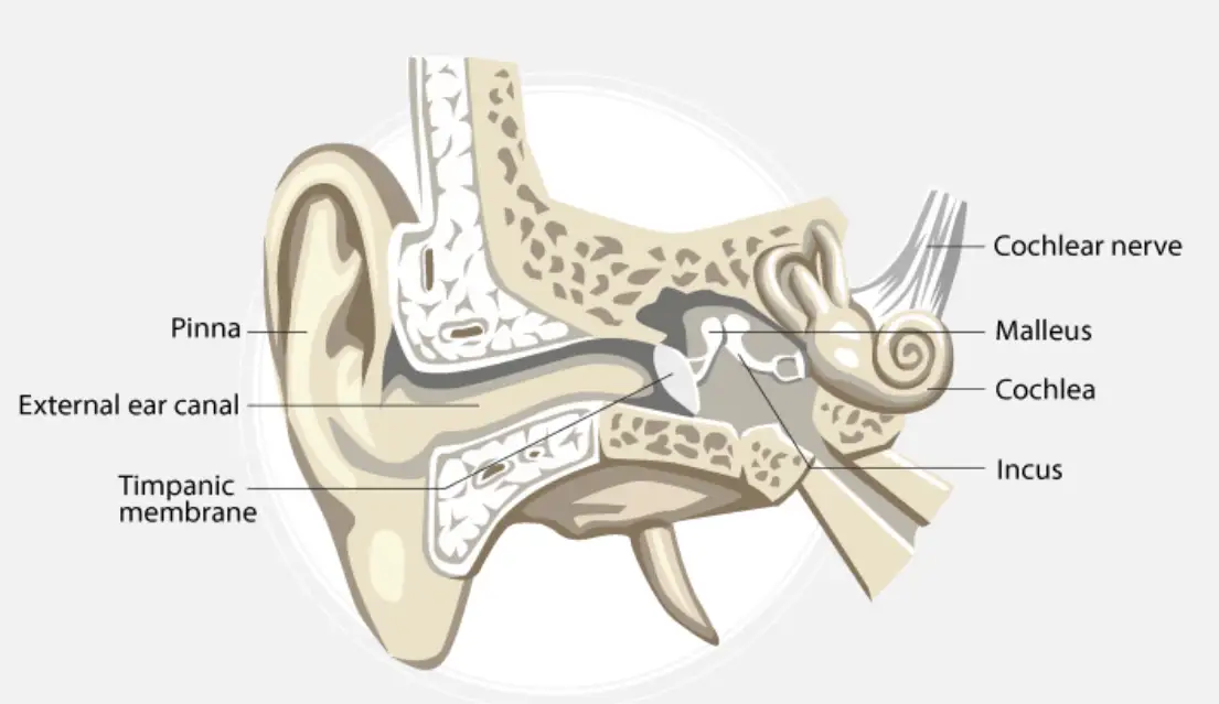
The external ear is composed of several distinct parts that work together to capture and channel sound waves efficiently. The auricle, also known as the pinna, is a flexible structure made of elastic cartilage covered by skin. Its curved shape acts like a funnel, gathering sound vibrations from the environment and directing them inward. The lobule, or earlobe, contains fatty and fibrous tissues richly supplied with blood vessels.
Extending inward from the auricle is the external auditory canal, a slightly curved passage supported by bone internally and cartilage externally. This canal is lined with stratified epithelial cells and contains ceruminous glands that produce earwax, which protects the ear from dust and microorganisms.
At the end of this canal lies the tympanic membrane, commonly called the eardrum. This thin, semi-transparent membrane separates the external ear from the middle ear and vibrates in response to incoming sound waves. The central depression of the membrane is termed the umbo, which plays a key role in sound transmission.
Illustration of the external ear structure
Middle Ear Structure and Its Functionality
The middle ear is an air-filled cavity situated between the external ear and the inner ear. It is bounded externally by the tympanic membrane and internally by a bony wall. This cavity contains the tympanic cavity and the auditory ossicles, which are essential for sound transmission.
The tympanic cavity connects to the nasopharynx through the eustachian tube, a canal approximately 3.5 cm long that helps equalize air pressure on both sides of the eardrum, ensuring optimal vibration.
Within the middle ear are three tiny bones called ossicles: the malleus, incus, and stapes. The malleus, shaped like a hammer, attaches directly to the tympanic membrane and connects to the incus. The incus resembles an anvil and links the malleus to the stapes. The stapes, the smallest bone in the human body, transmits vibrations from the incus to the oval window of the inner ear.
Inner Ear Composition and Sensory Functions
The inner ear consists of a complex bony labyrinth enclosing a membranous labyrinth. The bony labyrinth includes the vestibule, three semicircular canals, and the cochlea, all filled with a fluid called perilymph. Surrounding this is the membranous labyrinth, which contains endolymph and houses sensory receptors responsible for hearing and balance.
The membranous labyrinth features three semicircular ducts, the cochlear duct, saccule, and utricle. Sensory structures such as the organ of Corti (for hearing), cristae, ampullaris, and maculae (for balance) are embedded within these ducts and sacs.
Primary Functions of the Ear: Hearing and Balance
Process of Hearing: From Sound Waves to Brain Signals
Hearing begins when sound waves enter the external auditory canal and strike the tympanic membrane, causing it to vibrate. These vibrations are transmitted through the ossicles in the middle ear, which amplify the sound and convey it to the oval window of the cochlea in the inner ear.
Within the cochlea, the organ of Corti contains hair cells that convert mechanical vibrations into electrical impulses. These impulses travel via the auditory nerve to the brain, where they are interpreted as sound.
Mechanisms Maintaining Body Equilibrium
Balance is regulated by the vestibular system within the inner ear and the eustachian tube in the middle ear. The eustachian tube maintains equal air pressure on both sides of the tympanic membrane, which is crucial for proper ear function and balance.
The vestibular apparatus, including semicircular canals and otolith organs, contains receptors that detect head movements and position. These receptors send signals to the brain to help maintain posture and equilibrium.
Summary Table for Quick Revision
Ear Part | Structure | Primary Function |
|---|---|---|
Auricle (Pinna) | Elastic cartilage with skin covering | Collects and directs sound waves |
External Auditory Canal | Curved canal lined with epithelium and wax glands | Channels sound to tympanic membrane |
Tympanic Membrane | Thin membrane separating external and middle ear | Vibrates to transmit sound |
Ossicles (Malleus, Incus, Stapes) | Three small bones in middle ear | Amplify and transmit vibrations |
Eustachian Tube | Tube connecting middle ear to nasopharynx | Equalizes air pressure |
Bony Labyrinth | Vestibule, semicircular canals, cochlea filled with perilymph | Protects membranous labyrinth |
Membranous Labyrinth | Contains sensory receptors and endolymph | Detects sound and balance |
Organ of Corti | Hair cells inside cochlear duct | Converts vibrations to nerve impulses |
Vestibular Apparatus | Sensory receptors in semicircular ducts and otolith organs | Maintains equilibrium |
Glossary of Key Terms
Term | Definition |
|---|---|
Auricle (Pinna) | The external part of the ear that collects sound waves. |
Tympanic Membrane | A thin membrane that vibrates in response to sound waves. |
Ossicles | Three small bones (malleus, incus, stapes) transmitting sound vibrations. |
Eustachian Tube | Tube connecting middle ear to throat, equalizing pressure. |
Bony Labyrinth | Rigid, bony outer wall of the inner ear. |
Membranous Labyrinth | Fluid-filled tubes inside the bony labyrinth containing sensory cells. |
Organ of Corti | Structure in cochlea with hair cells that detect sound vibrations. |
Perilymph | Fluid filling the bony labyrinth. |
Endolymph | Fluid inside the membranous labyrinth. |
Vestibular Apparatus | Inner ear structures responsible for balance. |
Frequently Asked Questions
What is the role of the tympanic membrane in hearing?
The tympanic membrane vibrates when sound waves hit it, converting air vibrations into mechanical movements that are transmitted to the middle ear ossicles.
How do the ossicles amplify sound?
The ossicles act as a lever system, increasing the force of vibrations from the tympanic membrane to the oval window, enhancing sound transmission to the inner ear.
Why is the eustachian tube important for ear health?
It equalizes air pressure on both sides of the tympanic membrane, preventing damage and ensuring proper vibration for hearing and balance.
What sensory structures in the ear help maintain balance?
The semicircular canals, utricle, and saccule contain hair cells that detect head movements and position, sending signals to the brain to maintain equilibrium.
How does the organ of Corti convert vibrations into nerve impulses?
Hair cells in the organ of Corti bend in response to fluid vibrations, triggering electrical signals that are transmitted to the brain via the auditory nerve.